A woman, 62 years of age, presented to the emergency department (ED) with right-sided pleuritic chest pain and dyspnoea following a long road trip from Adelaide to Northern Queensland. In the morning of the presentation, she had a coughing fit and became acutely short of breath. On arrival at the ED, she was anxious but had normal observations (heart rate: 80 beats per minute, blood pressure: 114/65 mmHg, oxygen saturation: 96% on room air, Glasgow Coma Scale: 15, afebrile). She had no significant past medical history, was a non-smoker, had no regular medications and had a body mass index (BMI) of 22 kg/m2.
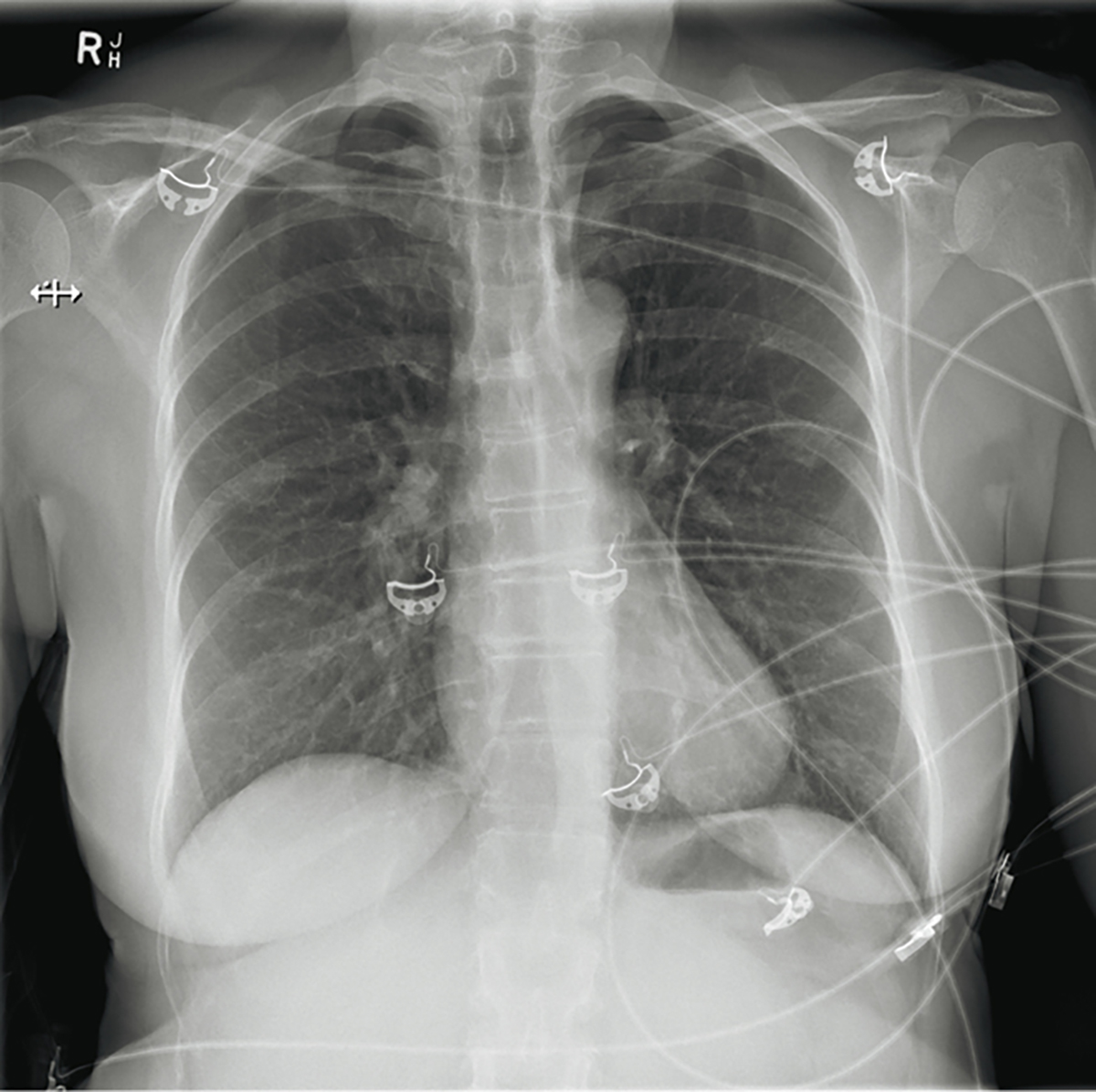
Chest auscultation was clear in all zones, heart sounds were dual with no murmurs and there were no signs of deep vein thrombosis. Given this, her Wells score totalled 1.5 (because of immobilisation while travelling), making pulmonary embolism (PE) unlikely and, therefore, a D-dimer test was undertaken to rule out a PE. Along with a D-dimer test, a troponin level was requested, an electrocardiogram (ECG) was performed and a chest X-ray (CXR) undertaken. The ECG was unremarkable, troponin was negative and D-dimer test was positive. The CXR is shown in Figure 1.

Figure 1. Anteroposterior chest X-ray
The X-ray shows loss of apical lung markings in keeping with pneumothorax
The CXR showed no signs of pulmonary embolism; however, it did reveal, rather surprisingly, bilateral pneumothoraces with no apparent cause. The patient had no prior history of lung disease, no history of pneumothraces and no connective tissue disorders. Further extensive examination revealed multiple small bruises in two parallel lines on either side of the patient’s spine, from her neck to lumbar region. On this discovery, she recalled that three days earlier she received acupuncture for neck pain.
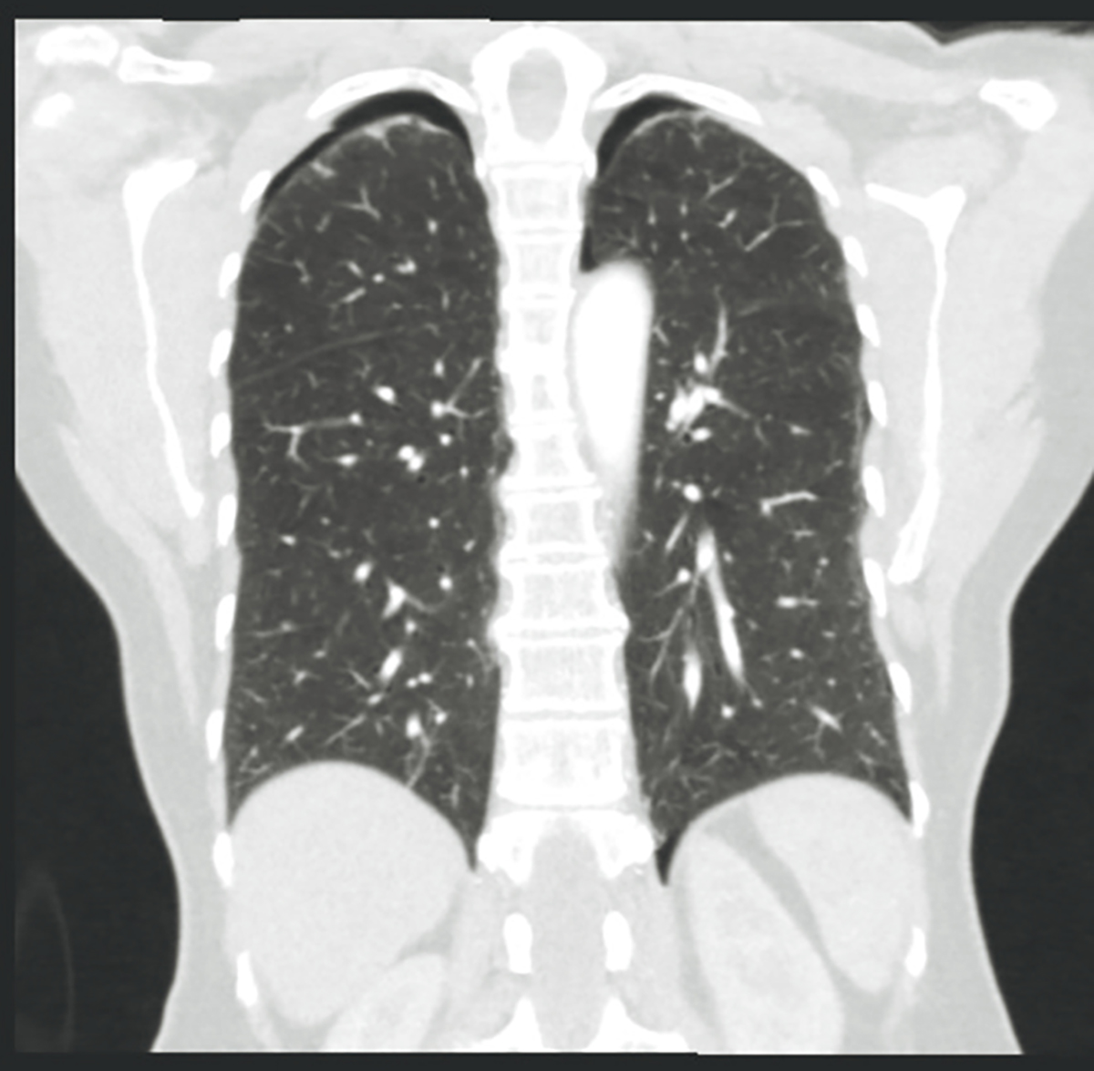
Computed tomography (CT) of the chest confirmed pneumothoraces and found no other underlying pathology (Figure 2).

Figure 2. Coronal CT lung scan
The scan shows small bilateral pneumothoraces
Thus, the diagnosis was traumatic, iatrogenic, bilateral pneumothoraces that were secondary to acupuncture. It is likely that at least one needle on either side of the spine pierced the visceral pleura, resulting in a slow air leak that accumulated over the three days. The leak on the right eventually caused pleuritic pain, shortness of breath, cough and anxiety that were severe enough to present to the ED.
The patient was admitted to the ward for monitoring over a four-day period without requiring an intercostal catheter, and serial CXR confirmed slow, steady resolution. She was discharged once she was stable, with a planned CXR in four weeks to ensure complete resolution.
Pneumothoraces from acupuncture, although rare, can occur when the needle pierces the parietal and visceral pleura, and air escapes from the lung into the pleural cavity.1 Given the fine gauge of the needle (0.12–0.35 mm), it is not uncommon for pneumothoraces to be delayed in presentation, as the rate of their development can be remarkably slow.
A literature review reveals that a pneumothorax is a very uncommon but serious complication of acupuncture. The rate of pneumothorax is estimated to be one to two events per 250,000 acupuncture treatments, and bilateral pneumothoraces are much rarer still.2,3
Factors increasing the risk of pneumothoraces may relate to the acupuncturist (needle punctures that are >20 mm, probably more likely with an inexperienced acupuncturist) or patient
(in thin patients, the lung surface can lie just 10–20 mm below the skin surface at the medial scapular area and midclavicular line).4
This case highlights that although acupuncture is one of the most commonly used alternative medicines, it is not without risks. These risks, being primarily acupuncturist-related, are mitigated in Australia by thorough education and training of registered practitioners. If patients are considering acupuncture, they should be informed about the risk of complications and all clinicians should be aware of possible signs and symptoms following an adverse outcome from such a practice.
Key points
- Acupuncture is not without risks; pneumothoracies can occur, albeit rarely.
- The prevalence of complications is higher when performed by inexperienced operators and in patients who are thin.
- If acupuncture is to be undertaken, a certified experienced practitioner is recommended to minimise risk of adverse effects.
Author
Joshua E Mann BSc, MBBS, RACGP Registrar GPT3, Royal Australian Air Force, RAAF Base Amberley, Amberley, Qld. mann.jo@hotmail.com
Competing interests: None.
Provenance and peer review: Not commissioned, externally peer reviewed.