The nose is the most prominent feature projecting from the face and is prone to injury arising from facial trauma. Injury can occur in all age groups and from a variety of causes, which may be either blunt or penetrating. The majority of injuries result in either bruising alone or a simple nasal fracture. Nasal fractures should be referred to ear, nose and throat (ENT) or maxillofacial services for prompt reduction (ideally one to two weeks from injury).
Complicated injuries include suspected facial fractures, full thickness lacerations, septal haematoma, septal abscess and cerebrospinal fluid (CSF) leak. It is critical that these are recognised early and managed appropriately in primary care
Anatomy
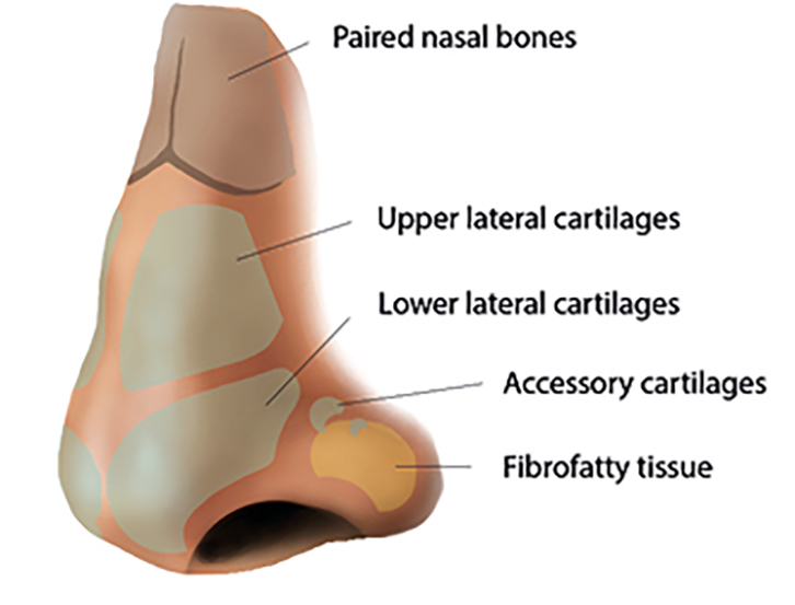
The nose can be subdivided into thirds. The upper third is made up of the paired nasal bones, which are attached to the frontal bones superiorly, forming a pyramid-shaped bony vault. The perpendicular plate of the ethmoid fuses with the nasal bones on the inner aspect, providing additional support. The middle third is composed of the quadrilateral cartilage of the septum in the midline and the upper lateral cartilages laterally. The lower third contains fibrofatty soft tissues of the nasal tip, supported by the lower lateral (alar) cartilages (Figure 1).

Figure 1. Anatomical illustration of nasal bones and cartilages
The nerve supply is from the ophthalmic and maxillary divisions of the trigeminal nerve. Blood supply to the nose has contributions from the internal and external carotid arteries. Venous drainage occurs anteriorly via the facial vein and posteriorly via the pharyngeal and pterygoid plexus. These pathways lack internal valves, creating the risk of retrograde intracranial infection.1
Uncomplicated nasal injuries
Nasal bones are the most commonly fractured facial bones.2 The mainstay of assessment is a thorough history and physical examination. Important aspects of history include injury mechanism, if there was immediate deformity (prior to soft tissue swelling), and new-onset nasal obstruction. It is also important to note any previous nasal injury or pre-existing deformity.
Physical examination involves assessing the degree of bony deformity by inspection and palpation, and excluding features of a complicated nasal injury.2 Soft tissue oedema arises within hours of injury and may significantly affect assessment.
Plain X-ray investigations are frequently inaccurate and generally do not contribute to the assessment of nasal injuries. False positive results can occur from previous injuries, prominent vascular markings or suture lines, whereas cartilaginous injuries can cause a false negative result.3
Cartilage deformity
If the nasal bones are midline but a cartilaginous septal deformity exists, these injuries are non-reducible acutely as the tissues spring back to their deformed state. It is appropriate to exclude a septal haematoma. If there is persisting nasal obstruction after one month of the injury, patients can be referred to ENT outpatient services for consideration of elective septoplasty.
Uncomplicated nasal fracture
Nasal fractures are generally managed with closed reduction under local or general anaesthesia. The choice of anaesthesia does not affect the success rate.4 Whenever possible, patients with suspected nasal fractures should be referred to an ENT service. Closed reduction should be performed once oedema resolves, ideally within 10–14 days of the injury.
In remote areas that do not have timely access to ENT services, appropriately skilled general practitioners (GPs) may perform closed reduction under local anaesthesia in a compliant adult patient. A local anaesthetic, such as lignocaine with adrenaline, is effective when administered into the root of the nose and lateral aspect of the bones.4 Severe injuries with gross external deformities or compound nasal fractures require early surgical intervention and should be referred to the emergency department immediately.2
Paediatric patients have incomplete ossification of nasal bones and a greater proportion of nasal cartilage; hence, they are prone to greenstick injuries.5 The ideal time frame for reduction is three to five days after the injury, and early referral to an ENT service is needed.6 In very frail patients, or patients with advanced dementia who have nasal fractures with only minor cosmetic change, it is prudent to discuss the option of leaving the fracture to unite, albeit with slight deformity remaining. This is often more appropriate to avoid further discomfort and potential harm from anaesthetic agents.
Complications of nasal injury
Lacerations
Traumatic lacerations of the nose are common. Debride and irrigate the wound well before closure.7 Most skin lacerations without involvement of cartilage or mucosa can be repaired with 6-0 non-absorbable monofilament sutures. Place sutures with small bites as nasal skin has little redundancy and poor flexibility.8 Refer deeper lacerations involving cartilage or mucosa to ENT or plastics services on the same day. Selected lacerations that are clean and have well-opposed edges may be closed with tissue adhesives, particularly in children.7 Determine and update tetanus immunisation status.
Epistaxis
Epistaxis is common with nasal injuries caused by trauma to the nasal mucosa and vessels. To administer first aid, the nose should be held firmly with the thumb and forefinger, closing the nostrils. The head is tilted forwards and the patient instructed to spit rather than swallow the blood. Continue pressure for 10–20 minutes.9 If available, vasoconstrictors such as cophenlycaine may slow or stop bleeding. Patients with haemodynamic instability or persistent bleeding (particularly patients taking anticoagulant agents) should be referred to the emergency department.
Nasal packing may be required to stabilise a patient for transfer. It is essential to ensure these are placed horizontally along the floor of the nose. Nasal packing should not be directed superiorly as patients with persistent bleeding may have associated nasoethmoid orbital fracture. In these circumstances, proper technique prevents intracranial placement of a nasal pack.
To facilitate nasal packing, there is a range of commercial products available, with Merocel and Rapid Rhinos commonly used in Australia. Merocel (Medtronic, Minneapolis) is a nasal tampon that expands with saline and is efficient for anterior packing.10 Rapid Rhino (AthroCare, Austin) is a balloon device available in sizes suitable for anterior and posterior packing.10 Rapid Rhinos are soaked in sterile water before insertion into the nasal cavity. Once inserted, the attached balloon can be inflated with air to increase the volume and pressure of packing. Rapid Rhinos are found to be more comfortable than Merocel.10
If commercial nasal packing is not available, anterior nasal packing can still be achieved with layered insertion of ribbon gauze soaked in petroleum jelly (Vaseline). Alternatively, intranasal insertion of cotton pledgets soaked in tranexamic acid has been shown to be effective treatment for anterior epistaxis.11 In suspected posterior epistaxis, a Foley catheter is advanced into the posterior oropharynx, then inflated. The anterior nasal cavity can then be packed with Vaseline gauze. This packing method can be secured in place with a clamp over the gauze.10
All patients with nasal packing should be placed on oral antibiotics (eg amoxicillin, cephalexin) to prevent toxic shock syndrome.10 These patients should be referred promptly to ENT services for admission as there is a risk of inadvertently dislodging the nasal pack into the oropharynx. The nasal packs are left in situ for three to five days to facilitate mucosal healing.10
Septal haematoma and abscess
Nasal septal haematomas occur as a significant complication of nasal trauma in 2% of nasal injuries.12 Blood vessels in the overlying mucoperichondrium supply the septal cartilage, which can be injured resulting in formation of septal haematoma. If left undrained, a septal haematoma can develop into a septal abscess or lead to ischaemic necrosis of the septal cartilage in a delayed manner and subsequent saddle nose deformity.12,13 Septal abscesses can result in meningitis, intracranial abscesses and cavernous sinus thrombosis because of the valveless venous drainage pathways creating an intracranial entry point.13
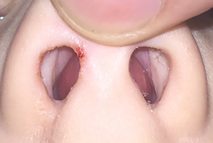
On examination, nasal septal haematomas show bilateral septal swelling obstructing the nasal airway, which is boggy on palpation with a blunt instrument (Figure 2). This is in contrast to a cartilaginous deformity (convex in one nostril, concave in other) with a firmness to palpation.
Septal abscesses are a delayed complication of nasal trauma. It is common to have progressive worsening bilateral nasal obstruction with increasing pain, headache and malaise. There may be fevers and purulent nasal discharge. Examination findings are similar to septal haematomas. Assessment should include vital signs and neurological examination.13 Nasal septal haematomas and abscesses require intravenous (IV) antibiotics and urgent drainage, and the patient should be referred to the emergency department.

Figure 2. Clinical photograph showing septal haematoma in a paediatric patient
Cerebrospinal fluid rhinorrhoea
The presence of thin, clear rhinorrhoea after nasal trauma should be considered CSF leak until proven otherwise. This can result from fracture and associated dural tear of the cribriform plate in the anterior skull base.9 The time frame for presentation can range from two days to three months post-injury.14 Typically, there is a positional element to the rhinorrhoea, occurring when patients lower their head forwards.
If CSF rhinorrhoea is suspected, a few drops of the fluid should be collected and sent for beta-2-transferrin assay, which is specific for CSF.14 These patients should be transferred immediately to a hospital with ENT and neurosurgery support for further management.
Nasal injury with facial fracture
Traumatic nasal injuries with high-force trauma should be suspected for concomitant facial fractures. Computed tomography (CT) imaging should be ordered and these patients referred to plastics or maxillofacial services for assessment if indicated. Table 1 provides a summary of associated facial fractures.
Table 1. Clinical features and assessment of facial fractures associated with traumatic nasal injuries15,16
|
Fracture type
|
Assessment essentials
|
Key assessment findings
|
Key points/management
|
|
Mandibular fracture
|
- Palpate mandible
- Inspect mandible dentition
- Assess mouth occlusion
|
- Trismus
- Malocclusion
- Chin numbness (mental nerve injury)
|
- Second most frequent fractured facial bone
- Angle and body most common fracture site
- Refer for CT facial bones and maxillofacial services (within 24 hours)
|
|
Zygomaticomaxillary complex fracture
|
- Palpate zygoma and maxilla
- Intraoral and intranasal examination
- Visual acuity and range of eye movement
- Mid-face sensation
|
- Mid-face numbness
- Malar depression
- Enopthalmus
- Trismus
- Malocclusion
|
- Fractures may involve lateral orbital wall, zygomatic arch, anterior or lateral maxillary sinus wall, or orbital floor
- Refer for CT facial bones and maxillofacial services (within 24 hours)
- Ophthalmology review for visual symptoms or orbital injury
|
|
Frontal fracture
|
- Palpate frontal bar
- Assess forehead sensation
- Visual acuity
- Assess for CSF leak
|
- Forehead lacerations
- Forehead numbness
- Epistaxis
- Rhinorrhoea
|
- Prone to injury due to anatomic position
- CT facial bones and sinuses
- Be wary of intracranial complications
- Delayed complications include CSF leak and frontal sinusitis
|
|
Orbital fracture
|
- Palpate orbital rims
- Examine eyelids and globe position
- Visual acuity and range of eye movement
- Forehead sensation
|
- Visual changes
- Forehead/mid-face numbness
- Enophthalmos
- Chemosis
Sub-conjunctival haemorrhage
|
- Essential to document visual acuity and range of eye movements
- CT facial bones and sinuses
- Ophthalmology review for visual symptoms or orbital injury (within 24 hours)
|
|
Nasoethmoid orbital fracture
|
- Palpate nasal bones
- Visual acuity
- Examine eyelids and globe position
- Palpate and exert pressure on medial orbital rim
|
- Posterior displacement of nasal pyramid
- Telecanthus
- Enophthalmos
- Epiphora
|
- Prone to injury in high-velocity mid-facial trauma
- Nasoethmoid orbital fractures can be minimally displaced
- Mobility or crepitus on palpation is abnormal
- Refer for CT facial bones and maxillofacial services (within 24 hours)
|
|
CSF, cerebrospinal fluid; CT, computed tomography
|
Conclusion
Traumatic nasal injuries encompass a wide range of potential complications, where prompt recognition and timely management are key to good functional and aesthetic outcomes.
Authors
Joo H Koh MBBS, DipSurg. Anatomy, ENT Service Registrar, Barwon Health, Geelong, Vic. joohoekoh@gmail.com
Osama Bhatti MBBS, General Practice Registrar, Southern GP Training, Corio Bay Medical Centre, Corio, Vic
Abbas Mahmood MBBS, FRACGP, GP Consultant, Training Supervisor – Southern GP Training, Corio Bay Medical Centre, Corio, Vic
Nicholas Agar MBBS, FRACS (OHNS), ENT Surgery Consultant, Barwon Health, Geelong, Vic
Competing interests: None.
Provenance and peer review: Not commissioned, externally peer reviewed.
References