The most common chronic wounds seen in general practice are venous ulcers, arterial ulcers, mixed aetiology ulcers (venous and arterial), pressure ulcers, skin tears and atypical wounds such as vasculitic ulcers. The management of ulcers is complex. Current practice varies in different clinical settings and treatments are often delivered by different healthcare professionals, who may or may not use best practice guidelines. The process described in this article is that used in the Therapeutic guidelines: Ulcer and wound management.1
The issue is not so much the choice of product but the accurate diagnosis of the underlying cause of the wound. It is often the case that a product is applied to a wound and when not successful the product is considered at fault so a different one is used; when that fails, the next product is used. The simple rule is to treat the whole patient and not the hole in the patient.
The three principles of wound management are:
- Define the aetiology.
- Control factors affecting healing.
- Select appropriate local environmental management (dressings).
Define aetiology
The most common chronic wounds seen in practice are:
- leg ulcers (venous, arterial, mixed)
- pressure wounds
- skin tears.
Venous leg ulcers
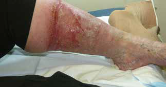 |
| Figure 1. Venous leg ulcer |
An estimated 400,000 Australians have venous leg ulcers (VLUs; Figure 1) due to chronic venous insufficiency (CVI). VLUs are managed in primary care or the community with variation in treatment and effectiveness,2 which in 2010 translated to healthcare costs of over $2 billion per year.3 The burden of recurrence is expected to rise with an ageing population and the growing epidemic of diabetes and obesity, which will further increase healthcare costs.4
VLUs result from the breakdown of the venous circulation in the leg and are associated with the inability of the leg to force the passage of blood through the various connecting veins via the bicuspid valves by muscular contraction. The increased venous pressure leads to pitting oedema, which in turn affects perfusion of the skin, so when some trauma occurs there is insufficient supply for healing to occur and an ulcer develops. Pitting oedema may result not only from chronic venous insufficiency but also organ failure, lymph disease or from medication (eg calcium channel blockers).
Venous ulcers commonly develop in the lower one-third of the leg (the gaiter area) and are usually irregular in shape. Pitting oedema is usually present. The skin is often stained around the ulcer area because of haemosiderin deposition after leakage of red blood cells from the circulation. Typical features of venous ulcers include skin changes such as eczema or atrophy blanche (white stippled scars on the skin). The three most common risk factors for VLUs are a history of obesity, past deep vein thrombosis (DVT) and poor mobility resulting in venous stasis.3,5
In some cases, treatment includes surgery; however, the mainstay of treatment is the application of graduated compression therapy toe-to-knee (30–40 mmHg at the ankle). It is, however, essential to exclude arterial involvement by testing the ankle brachial index or by ultrasonography. Lower limb exercise and addressing occupational factors, such as long periods of standing leading to venous stasis, should be encouraged.
It is important to keep in mind that oedema may result not only from venous disease (pitting oedema), but also other causes including organ failure, lymph disease or from medication (eg calcium channel blockers). Lymphoedema is caused by a reduction in the function of the lymph vessels to drain extracellular fluid. The resultant oedema will place the patient at risk of ulcer development as a result of minor trauma and by the hyperkeratotic nature of the skin.6
Arterial ulcers
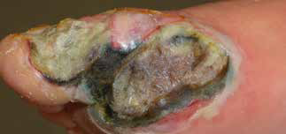 |
| Figure 2. Arterial ulcers |
The death of skin automatically follows occlusion of its arterial blood supply unless this is gradual enough to allow a collateral blood supply to be established. Atheroma (thickening) is the most common cause of arterial ulcers of an ischaemic nature.
Ischaemic pain, especially at night, is associated with arterial ulcers (Figure 2). This is as marked in small ulcers as in larger ulcers. Their edges are often sharply defined and the ulcer is ‘punched out’. The base is often covered with slough. This may deepen to expose tendons. A history of intermittent claudication (pain on exercise), dependent foot (dusky foot) white on elevation, peripheral vascular disease, lower ankle brachial pressure index, weak/absent pulses, sluggish/poor capillary refilling may be present. Commonly, the ulcer site is below the ankles and on the foot or toes; however, arterial ulcers may be present on other areas of the body. The skin is often shiny and friable. Poorly controlled diabetes and smoking are significant risk factors causing arterial insufficiency.7
Treatment of arterial ulcers may involve surgical intervention for angioplasty, stenting, bypass grafting and, ultimately, amputation. Pain control is an important aspect of the management of arterial ulcers. Adequate analgesia is required to manage the severe ischaemic pain often experienced with arterial ulcers. These wounds should not have compression applied even if there is some associated venous disease.7
Venous/arterial or mixed ulcers
It is important to note that 15–20% of leg ulcers are of mixed aetiology. These ulcers are often difficult to heal because of associated oedema, cellulitis, thrombophlebitis, rheumatoid diseases, particularly in patients who are bedridden, and malnourishment-related conditions of the skin in elderly patients. The most important issue is to determine whether the predominant cause is venous or arterial and then treat it. Graduated compression may be contraindicated, depending on the extent of the arterial component of the problem. If compression cannot be used it is difficult to address the venous component of the problem.1,8,9
Skin tears
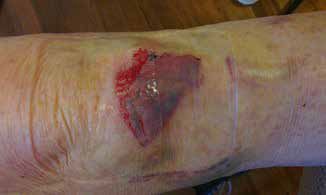 |
| Figure 3. Skin tear |
Skin tears (Figure 3) are the most common wound type in the elderly population. If treated inappropriately, skin tears can become chronic wounds, exerting huge costs on the community and deleterious effects on the individual’s physical and psychological health.
The ageing process will affect most of the structures of skin through loss of hair follicles, sebaceous glands that supply natural moisture to the skin, receptors, blood supply and sweat glands. The result of these tissue changes is that the skin becomes thinner and brittle, and the blood supply is reduced, fragile and more prone to injury. It is critical to identify patients at risk and then introduce prevention strategies. A recent study in Western Australia of 900 patients in 23 nursing homes showed a 50% reduction in skin tears and a significant cost saving by applying a moisturising lotion twice daily.10
The management of a skin tear will depend on the level of damage. If it is possible to replace the flap, this should be done carefully, holding it in place with a few adhesive strips applied with no tension and covering with a silicone foam dressing, then covering with one or two layers of tubular compression bandages to apply mild pressure on the wound. This system is reviewed after 3 days then redressed every 5–7 days until the wound has healed.1,11
In addition to the more common forms of ulceration, there are a number of less common causes. Vasculitic ulcers may develop as a result of a number of medical conditions, such as rheumatoid arthritis and polyarthritis, which cause damage to the microarterial circulation by circulating antibodies. These wounds are often misdiagnosed as venous ulcers. If there is a dark wound margin, purple discolouration in the peri-skin and the wound is painful, suspect vasculitis. These wounds need to be treated systemically as well as topically.1,12
Pressure ulcers
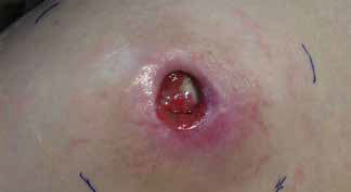 |
| Figure 4. Pressure wound |
Pressure ulcers (Figure 4) are the most preventable of all of the chronic wounds.
Patients who are bedridden, for example, as a result of stroke, spinal injury, multiple sclerosis or dementia, often develop extensive pressure ulcers. These wounds may result from direct pressure, friction or shear injuries.
A pressure wound develops when capillary blood flow to the skin and tissue over a bony prominence is decreased for a sufficient period of time. The consequence of the restricted blood supply is a reduction in oxygen supply and nutrition to the tissue, and inadequate excretion of the waste products.
Friction
Friction occurs when the top layers of skin are worn away by continued rubbing against an external surface. This can be caused by ill-fitting footwear, or even bed linen, and can manifest in a simple blister or tissue oedema, or an open pressure wound.
Shear
Shear occurs when the skin remains in place, usually unable to move against the surface it is in contact with, while the underlying bone and tissue are forced to move. This force will contribute to the destruction of microvasculature in a manner similar as direct pressure.
The management of a pressure wounds requires the removal of all pressure on the wound, increased nutrition and, depending on the size and depth of the wounds, the use of topical or cavity products.1,13
Control factors that affect healing
Most wounds heal readily but others are slow to heal or remain unhealed for a considerable length of time. A number of factors, including intrinsic and extrinsic factors,14,15 affect wound healing (Table 1). These should be considered and addressed as part of a holistic approach to wound management.
Table 1. Intrinsic and extrinsic factors affecting wound healing
|
| Intrinsic factors | Extrinsic factors |
|---|
Health status
- Good arterial and venous circulation: anaemia impairs oxygen transport
|
Mechanical stress
- Pressure
- Friction
- Shearing forces
|
Immune function
- Normal immune function helps to cleanse the wound
- Reduced function increases the risk of infection
|
Debris
- Slough
- Necrotic tissue
- Eschar
- Scab
- Dressing residue
- Sutures
|
Comorbidities
- Diabetes
- Rheumatoid arthritis
- Other diseases
|
Dessication
- Drying of the wound surface results in death of surface cells
|
Age-related changes to skin
- Loss of hair follicles, sebaceous glands, receptors
- Reduced blood supply
- Increased fragility
- Dryness
- Thinning
|
Maceration
- Excess moisture retards healing and damages the
peri-skin
Temperature
|
Nutrition
- Balanced diet including proteins (particularly for the amino acid arginine), carbohydrates, fats and fluids promotes healing
|
Infection
Chemical stress may have an adverse effect on the wound and cells
- Topical agents (eg antiseptics)
- Smoking20
- Drugs (eg steroids and non-steroidal anti-inflammatory drugs)21–23
|
Wound management
The management of the wound environment is now based on the concept of wound bed preparation (WBP) interventions, which address debridement, bacterial balance, exudate management and the local tissue in the wound environment. These important assessment elements have led to the development of the concept of the TIME principles (Tissue, Inflammation/Infection, Moisture, Edge/Epithelialisation), overseen by the World Union of Wound Healing Societies.1,16,17 The Therapeutic guidelines: Ulcer and wound management uses this approach in guiding wound care in a best practice, evidence-based context.1
Much of the focus in wound management is on the dressing when, in fact, this is not the important aspect to address. It is often the case that a product is applied to a wound and when not successful the product is considered to be at fault so a different one is used and when that fails the next product is used. In general, the dressing does not heal the wound, but appropriate contemporary dressings facilitate the optimal environment to enhance wound healing. It is critical to first determine and address the underlying cause of the wound. The next step is to identify and address those factors affecting healing over which there may be some influence. Finally, the local wound management issues should be considered and dressing selection based on the function of the dressing (eg exudate management, debriding, cavity filler) and what is needed to optimise the local wound environment for healing. Once a venous ulcer is healed then consider the ongoing use of compression stockings for life.1,3,5
Wound management products
Passive dressings
For many years the products used were of the ‘passive’ or the ‘plug and conceal’ concept, including gauze, lint, non-stick dressings and tulle dressings. These products fulfill very few of the properties of an ideal dressing and have very limited, if any, use as primary dressing, but some are useful as secondary dressings.1,18,19
Interactive dressings
These dressings help to control the micro-environment by combining with the exudate to form either a hydrophilic gel or, by means of semipermeable membranes, controlling the flow of exudate from the wound into the dressing. They may also stimulate activity in the healing cascade and speed up the healing process.
There are six classes of interactive dressings, classified according to their functionality:1,18,19
- Film dressings
- Hydroactive dressings
- Hydrocolloid dressings
- Hydrogel dressings
- Foam dressings
- Alginate absorbent fibre dressings.
The choice of dressing will depend on the wound type and depth, level of exudate and the presence of bacteria. A comprehensive overview of wound dressings may be found in Wound Dressing Products Update.18 A wound identification and products selection guide can be found on the Department of Veterans Affairs website (see Resources).
Bandages
The use of material to bind the wound is as ancient as medicine itself. Techniques and material have changed little over the centuries, but in the past 15 years there has been an explosion in the types of bandages available. When choosing and applying a bandage it is important to differentiate between the traditional and the ritual on one hand and what is best and most cost-effective for the patient on the other.11
The bandage may be needed for several reasons:
- Retention: keeping a dressing in place
- Musculoskeletal support: supporting an injured joint
- Compression: assisting venous return from the lower leg.
Competing interests: Geoff Sussman has received payment for lectures at Ansmed Conferences.
Provenance and peer review: Commissioned, external review.
Resources
Department of Veterans Affairs. Wound identification and dressing selection chart resources. Canberra: Commonwealth of Australia; 2012. Available at www.dva.gov.au/service_providers/resources/Pages/WoundCareFlashVideo.aspx [Accessed 5 August 2014].