Tropical ulcers are relatively rare in Australia and can be challenging to diagnose. Clinical presentations include swellings, nodules, papules, abscesses and sinuses. They are best managed in collaboration with a dermatologist or infectious diseases physician. This article will discuss the important causes of tropical and exotic ulcers occasionally seen in Australia.
Tropical ulcers result from diseases transmitted by mosquitoes and flies in tropical and subtropical countries where the weather is hot and humid. Predisposing factors include poor hygiene, low socioeconomic conditions, poor nutrition, poverty and lack of protection from insect bites. Superinfection with Fusobacterium species, anaerobic microorganisms (Bacillus fusiformis) and spirochetes (Treponema vincenti) aggravates these ulcers and delays healing.1 The rising numbers of tourists, especially eco-tourism, visiting rural areas, workers or foreign aid workers and military personnel returning from tropical regions, as well as in immigrants from these countries have led to increasing numbers of tropical ulcers being seen in metropolitan Australian medical practice.
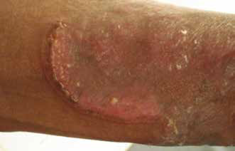
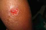
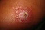
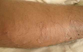
Typical presentation of a simple bacterial tropical ulcer
The lesion first appears as a small papule on the lower leg, usually at the site of trauma. The ulcer then spreads rapidly, forms a blister and undergoes necrosis, which is associated with pain, fever and malaise. Within a few weeks the ulcer enlarges to 10–12 cm and a superficial, necrotic, malodorous, purulent, haemorrhagic black coating develops. After 3–4 weeks, the edge becomes flatter, and the swelling and pain decrease.2
Simple tropical ulcers should be differentiated from other causes of leg ulceration that may be seen in the tropics. In contrast to buruli ulcers, for example, tropical ulcers show little undermining, whereas ulcers of cutaneous leishmaniasis are not usually painful. Appropriate investigations and skin biopsies may be needed to differentiate a tropical ulcer from other causes of chronic leg ulceration such as syphilitic gumma, pyoderma gangrenosum, venous ulcers haemoglobinopaties, vasculitic and neoplastic ulcers.
First-line treatment for tropical ulcers is a combination of penicillin (500 mg every 6 hours for 1 week) and metronidazole (250 mg every 8 hours for 10 days). For patients who are allergic to penicillin, tetracycline (minocycline 100 mg twice daily or doxycycline 100 mg twice daily) can be given instead. Antibiotic treatment should be combined with proper would care and cleansing, immobilisation and nutritional support. Large ulcers (>5 cm) require wound debridement and skin grafting
.
Other bacterial skin ulcers
In Australia, tropical bacterial infections include may include standard infections such as impetigo, pyodermas, folliculitis, boils, erysipelas and cellulitis, and can present within a few days of return from travel.
Blastomycosis-like pyoderma
This is a unique condition caused by Staphylococcus aureus and Pseudomonas species. It presents with large verrucous plaques, multiple pustules and draining sinuses. Blastomycosis-like pyoderma can resemble coral reef granuloma, a condition seen in actinically damaged skin of the elderly in subtropical areas of Australia.
Cutaneous anthrax
Cutaneous anthrax develops 1–7 days after exposure of lacerations to anthrax-infected sick animals or contaminated wool/fur. Infection gives rise to a pruritic papule that ulcerates and then forms a necrotic eschar over a few days.3
Mycobacterial skin ulcers
The clinical presentation of mycobacterial infections is influenced by the route of inoculation and host immunity. Table 1 outlines the types of cutaneous tuberculosis that cause exotic ulcers in Australia. Examples of these ulcers are shown in Figures 1 and 2.
Table 1. Cutaneous mycobacterial infections
|
|
Disease
|
Route of infection
|
Host immunity status
|
Features
|
Clinical mimics
|
|---|
|
Lupus vulgaris
|
Exogenous Haematogenous
|
Moderate-high-degree immunity
|
- Usually a solitary lesion
- Common in head and neck
- Initially reddish brown plaque with soft gelatinous consistency
- Diascopy shows apple jelly nodules
- Spreads peripherally with elevated infiltrated edges and central atrophy (Figure 1a, b)
- Classic plaque
- Keratotic, hypertrophic, ulcerative and vegetating forms seen
- Chronic large lesions can damage underling tissues and has an increased risk of squamous cancer formation
|
- Sarcoidosis
- Discoid lupus
- Deep fungal infections
- Lupoid leishmaniasis
- Basal cell carcinoma
- Pyoderma gangrenosum
|
|
Tuberculoid chancre
|
Exogenous
|
No previous immunity against
M. tuberculosis
|
- Develops at the inoculation site as a brownish papule and evolves into a firm, shallow, non-tender, undermined ulcer with a granulomatous base.
- Painless, regional lymphadenopathy is frequently seen
|
- Blastomycosis
- Histoplasmosis
- Coccidioidomycosis
- Sporotrichosis
- Tularemia
- Cat scratch disease
- Pyoderma gangrenosum
- Cutaneous pseudolymphoma
- Sarcoidosis
- Discoid lupus erythematosus
- Squamous cell carcinoma
|
|
Scrofuloderma
|
Endogenous – contiguous
|
Poor immunity against
M. tuberculosis
|
- Firm, painless, red-brown nodules develop over an infected gland or joint and suppurate and break down forming ulcers and multiple sinus tracts.
- Fibrosis and irregular scarring can occur
|
- Sporotrichosis
- Coccidioidomycosis
- Actinomycosis
- Chronic bacterial osteomyelitis
- Hidradenitis suppurativa
- Severe acne conglobata
|
|
Verrucous (warty) TB
|
Exogenous – direct inoculation
|
Previously immune
|
- Painless, solitary lesion at exposed sites
- Purplish or brownish warty nodule
- Painless
- Extends peripherally causing central atrophy
- Exudes pus or keratinous material
|
- Verrucose leishmaniasis
- Blastomycosis
- Chromoblastomycosis
- Sporotrichosis
- Verrucose tertiary syphilis
- Mycobacterium marinum
- Hypertrophic lichen planus
- Squamous cell carcinoma
- Psoriasis
|
|
Erythema induratum
|
Endogenous
– tuberculoid
|
Moderate-high-degree immunity
|
- Panniculitis presenting with multiple, painful, recurring, ulcerated nodules in the lower limbs.
- Common in women
|
- Nodular vasculitis
- Erythema nodosum
|
|
Papulonecrotic tuberculosis
|
Endogenous
– tuberculoid
|
Moderate-high-degree immunity
|
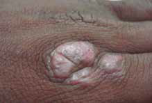
- Recurrent crops of hard red papules that ulcerate and leave scars (Figure 2)
|
- Pityriasis lichenoides
- Nodular prurigo
|
 |
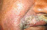
 |
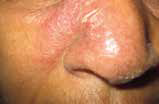
| Figure 1a. Lupus vulgaris in the arm with verrucous border and atrophic center |
Figure 1b. Lupus vulgaris lesion on the face |
Mycobacterial ulcers may resemble many other dermatological conditions and microbiological confirmation remains difficult. Clinical suspicion and awareness of the many clinical variants of cutaneous tuberculosis are required to avoid delays in diagnosis and treatment.
Diagnosis requires two skin biopsies. One should be sent in formalin for routine histology and Ziehl-Neelson staining. The other biopsy should be sent fresh (in saline-soaked gauze to prevent drying) for bacterial and mycobacterial culture and polymerase chain reaction (PCR). Typical histology includes caseating epithelioid granulomas that contain acid-fast bacilli. Mycobacterial culture has high specificity and low sensitivity and takes 4–6 weeksbefore results are available.4
 |
| Figure 2. Papular necrotic TB with ulcerative nodules and scarring |
PCR, which can be performed with small tissue samples,5 has higher sensitivity than culture, and results are available within a week. The interferon gamma release assay (QuantiFERON-Gold In-Tube test or T-SPOT TB test) has high sensitivity and specificity and can be used in latent and active tuberculosis. This test has largely replaced the Mantoux test as it has the advantage of lack of cross-reactivity to the BCG vaccine.6
In 10–20% of cases, the tests may fail to confirm presence of tuberculosis and in such cases, a trial of anti-tuberculosis treatment will be necessary.7 All forms of cutaneous tuberculosis are treated with a combination of antitubercular drugs, including isoniazid, rifampicin, pyrazinamide and ethambutol, over a period of several months; the dose regime is dependent on local protocols.
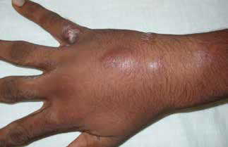
Atypical mycobacteria
Atypical or non-tuberculous mycobacteria, which are different from Mycobacterium tuberculosis, may contaminate soil or water. These bacteria cause a spectrum of infections ranging from pulmonary infection to papules, nodules, ulcers and abscesses in the skin.8 They cause opportunistic infections in immunocompromised patients, and skin lesions in healthy people following infection at sites of trauma.
Fish tank granuloma/swimming pool granuloma
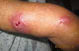
 |
| Figure 3a. Fish tank granuloma with sporotrichoid spread and lymphaedema |
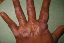
Fish tank granuloma is caused by M. marinum infection. It is most commonly seen in fishermen, oyster workers, swimmers and aquarium workers, and usually involves the dominant hand. The infection starts as a solitary papule or nodule, which grows slowly over several months. Induration and ulceration occur later. Sporotrichoid spread and lymphangitis occur subsequently9 (Figure 3a, b).
Diagnosis of atypical mycobacterial ulcers is similar to that of cutaneous tuberculosis. Two skin biopsies are required: a formalin-fixed sample for routine histology and Ziehl-Neelson staining, and a fresh sample in saline-soaked gauze for culture and PCR. The optimal temperature for atypical mycobacteria cultures is 30°C and laboratory personnel should be notified of this requirement. PCR can be used to detect M. marinum infection directly from the biopsy sample.
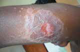
 |
 |
 |
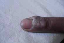
| Figure 3b. Fish tank granuloma – different clinical presentations |
Antibiotics are the mainstay of treatment and can be given as single agents (minocycline 100 mg twice daily, doxycycline 100 mg twice daily or clarithromycin 500 mg twice daily) or as combinations (minocycline + clarithromycin, minocycline + rifampicin or clarithromycin + rifampicin). Treatment should be continued for 1–2 months after resolution of visible lesions. Treatment duration is typically 3–6 months or longer if deeper structures are involved.10
Bairnsdale ulcer (Buruli ulcer)
Buruli ulcer is known in Australia as the Bairnsdale ulcer and is caused by M. ulcerans, an organism that is closely related to M. tuberculosis. It grows best at low oxygen concentrations and is found in muddy or marshy ground. It causes necrotising ulcers in otherwise healthy people. Buruli ulcer is found in many parts of Africa – the name is derived from Buruli, a province in Uganda. It has been also reported in other parts of the world including Papua New Guinea, Australia, China, India and South America.11
Acid-fast bacilli may be detected in a deep-tissue biopsies and this is helpful in differentiating Buruli ulcers from other causes of chronic leg ulceration. M. ulcerans can be cultured from ulcer exudates or from fresh tissue samples.
Oral rifampicin (10 mg/kg daily) combined with either clarithromycin (7.5 mg/kg twice daily) or a fluoroquinolone (moxifloxacin 400 mg daily or ciprofloxacin 500 mg twice daily) as a second oral agent is recommended.12 Antibiotics should be administered for 8–12 weeks. After 4 weeks of antibiotic therapy, surgery aimed at definitive wound closure may be considered.
Other non-tuberculous mycobacteria that can cause abscesses and ulcers include M. avium intracellulare (in patients with acquired immunodeficiency), M. szulgai, M. terrae, M. fortuitum, M. chelonae, M. malmoense, M. xenopi and M. abscessus.
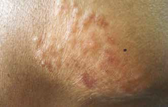
Cutaneous leishmaniasis
Leishmaniasis is caused by the parasitic protozoan Leishmania, which is transmitted by sandflies, a tiny sand-coloured blood-feeding fly that breeds in forest areas, caves and burrows in tropical and subtropical regions. There are several clinical variants of leishmaniasis, caused by different species of Leishmania. Cutaneous leishmaniasis is the most common form of the disease and is commonly seen in Iran, Afghanistan, Syria, Saudi Arabia, Peru, Brazil, the Middle East, North Africa and Asia. L. tropica, L. major, L. infantum and L. aethiopica cause old world cutaneous leishmaniasis, and L. tropica mexicana, L. braziliensis and L. amazonensis cause new world leismaniasis. Cutaneous leishmaniasis is a common cause of skin ulceration in travellers returning from these countries.
Clinical features of cutaneous leishmaniasis can vary. Typically, the lesion starts as a small red asymptomatic papule, which grows slowly into a plaque or nodule and ulcerates. There is a central crater covered with a dry scab with raised edges. There may be a halo around the lesion (Figure 4).
 |
 |
 |
 |
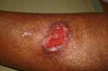 |
 |
 |
 |
| Figure 4. Different clinical types of cutaneous leishmaniasis |
In endemic areas, diagnosis of cutaneous leishmaniasis is usually made on clinical grounds alone. Microscopy of a slit skin smear or skin biopsy shows organisms within macrophages (dot and dash appearance). PCR and parasitic culture can be used to confirm the clinical diagnosis.
Small lesions of cutaneous leishmaniasis may resolve spontaneously over time and leave a visible scar. Intralesional antiparasitic pentavalent antimonials, such as sodium stibogluconate or meglumine antimoniate are the gold standard of treatment. Several injections given weekly are usually required until the lesions heal. A 7% solution of hypertonic saline or liquid nitrogen cryotherapy may also be given.13 Oral fluconasole and lipsosomal amphotericin B treatments have also shown promising results in case studies.14 As there is no vaccine currently available, travellers to endemic areas should take necessary preventive measures to avoid sandfly bites.
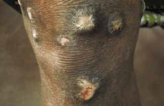
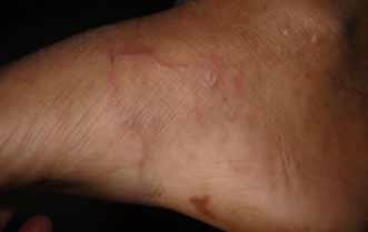
Cutaneous larva migrans
Cutaneous larva migrans is the most common tropically acquired dermatosis in travellers returning to Australia. Lesions are usually acquired by sunbathing or walking barefoot on sandy beaches contaminated by dog and cat faeces containing nematode larvae, which penetrate the skin. Ancylostoma braziliense (hookworm of wild and domestic dogs and cats) is the most common cause.15 Distal lower extremities, the dorsum of the feet and interdigital spaces of the toes are more commonly affected than other areas. Migration of the larvae causes itchy erythematous, serpiginous, cutaneous eruptions. Multiple infections may occur (Figure 5).
Albendazole 400 mg daily for 3 days is the treatment of choice. Ivermectin 200 µg/kg in a single dose is another treatment option. Antibiotics may be indicated for secondary bacterial superinfection. Liquid nitrogen cryotherapy of the advancing edge of larval burrow can also be used.
 |
 |
| Figure 5. Single and multiple lesions of cutaneous leishmaniasis |
Tropical mycoses
Tropical fungal infections, such as chromomycosis, sporitrichosis and mycetoma, can present as chronic skin nodules and ulceration.
Conclusion
Increasing international travel for tourism and employment, as well as immigration from tropical and subtropical countries, has led to an increasing number of tropical and exotic skin ulcers being seen in contemporary Australian medical practice. These ulcers may present as papules and nodules, vesicles and bullae, plaques, creeping dermatitis and cutaneous ulcers. The spectrum of dermatoses include infectious skin diseases (bacterial, mycobacterial, atypical mycobacterial, protozoan, nematode and tropical mycoses), as well as environmental causes such as arthropod-related reactions, contact dermatitis and marine-life dermatitis. Early recognition and treatment of these entities is facilitated by an increased awareness among primary care physicians and collaboration with the medical specialists.
Authors
Deepani Rathnayake MBBS, MD, Dermatology Fellow, Sinclair Dermatology, East Melbourne, VIC.
Rodney Sinclair MBBS, MD, FACD, Director and Head, Sinclair Dermatology, East Melbourne, VIC.
Competing interests: Rodney Sinclair has received payment from Bayer Healthcare and Leo Pharma for consultancy work, lectures and presentations, which are not related to this publication. Provenance and peer review: Commissioned, externally peer reviewed.
- Bowman DD, Montgomery SP, Zajac AM, Eberhard ML, Kazacos KR. Hookworms of dogs and cats as agents of cutaneous larva migrans. Trends Parasitol 2010;26:162–67.