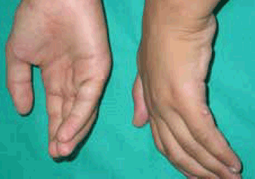
Figure 1. Soft, pink, asymptomatic and congenital papules in ulnar surface of proximal phalanx of the fifth fingers
Question 1
What is the differential diagnosis?
Question 2
What is the most likely diagnosis?
Question 3
How many types of this condition are we likely to find in our daily practice?
Question 4
Is it necessary to rule out any extra-cutaneous disease?
Question 5
What are the treatment options for this disease?
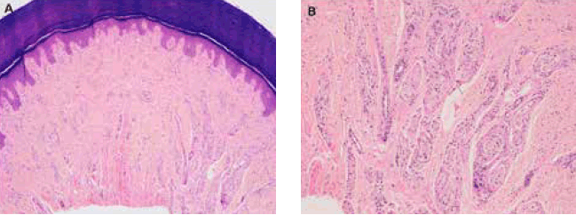
Figure 2. A. Hematoxylin-eosin staining showed the presence of a small wellcircumscribed dermal nodular tumor on fifth finger. B. Dermal proliferation of nerves and Meissner corpuscles
Answer 1
The main differential diagnosis is common warts, dermatofibroma, neurofibroma and dermal naevus.
Common warts are clinically hyperkeratotic and dome-shaped papules with punctate black dots that represent thrombosed capillaries. Dermatofibroma are hyperpigmented papules, sometimes normocoloured, minimally elevated and they may seem attached to subcutaneous tissue. Neurofibromas are skin-colored papules, usually solitary and soft. Dermal nevus feels soft to the touch. They could be hypopigmented or normocoloured but rarely are located on fingers. None of these entities are congenital.
Answer 2
Ulnar type B polydactyly.
Answer 3
All classifications have been performed with academic purposes. Swanson1 and Buck-Gramcko2 have classified polydactyly as radial/tibial affecting the thumb or great toe. When the affected digit is the little finger, the polydactyly is referred to as ulnar/fibular and cases in which the three central digits are affected are referred to as central polydactyly, the least common of the three types. This classification has replaced previous classifications into preaxial and postaxial and polydactylies. This has the advantage of avoiding confusion between the central morphological axis in the human hand, running along the third digit, and the more posterior metapterygial axis.3 Temtamy and McKusick4 prefers to divide polydactyly on type-A, when it is fully developed and type-B when it is not, including bilateral cases as the present case. A radiological study may help discern whether there is an associated skeletal component.
Answer 4
This is the most frequent congenital hand anomaly in people of African descent and the second most common, after syndactyly, in those of Caucasian descent. Isolated polydactyly is frequently inherited as an autosomal dominant trait of variable expression and its prevalence is approximately 1 in 531 live births. Although it often appears as a bilateral isolated feature usually underdiagnosed and misdiagnosed, an association with other birth defects such as mental retardation, underdevelopment, craniofacial dysmorphism, type 1 neurofibromatosis, hypospadias or hydroureteronephrosis is present in 6.6% of cases.5
Answer 5
Ulnar type B polydactyly is often successfully treated using suture ligation in the delivery room with absorbable sutures6 or surgery. We consider excision with high ligation of the accompanying digital accessory nerve (digital nerve in the supernumerary digit) to be safer and more effective. It avoids the risk associated with anesthesia for the newborn and the possible occurrence of post-surgical neuromas.7 Other ablative techniques such as shaving and cautery, cryotherapy or CO2 lasers have also been used.
Competing interests: None.
Provenance and peer review: Not commissioned; externally peer reviewed.