What is ionising radiation?
Ionising radiation is a form of electromagnetic radiation with enough energy to dislodge outer shell electrons from an atom, causing that atom to become ionised and charged.1 In practice, ionising radiation for medical imaging includes X-ray, CT, fluoroscopy and angiography. Ultrasound and magnetic resonance imaging (MRI) do not use ionising radiation.
How is ionising radiation harmful?
An ionised atom can cause production of free radicals, break or produce new chemical bonds, or damage molecules that control cell processes, such as DNA, RNA and cell proteins.2 These processes can result in cell death, repair or mutation. Mutation can lead to carcinogenesis. Cells that are dividing or least specialised are most vulnerable.2
How is harm due to radiation exposure measured?
Everyone is exposed to natural radiation, which varies regionally. Biological risk from radiation of any source is expressed as effective dose, and is measured in millisieverts (mSv). Effective dose is an estimation of potential harm from cancer and hereditary effects, and reflects both the energy of radiation and sensitivity of individual organs.3
The relationship between radiation dose and risk of adverse biological effect is assumed to be a linear, no-threshold model, but risk of harm is also related to dose rate, age at exposure, radiation type and organ exposed. Table 1 shows the typical effective doses of common X-ray, CT and nuclear medicine procedures. Effective doses vary relatively minimally across studies, and are changed periodically, as the International Commission on Radiological Protection updates organ weighting according to latest available estimates, eg. most recent estimates have increased weighting for the breast such that the effective dose for a chest CT has increased by 20%.3
Table 1. Comparative effective dose of common medical imaging procedures using ionising radiation
| Test | Effective dose (mSv) |
|---|
| X-ray |
|---|
| Extremity |
0.001 |
| Duel energy X-ray absorptiometry (DXA) |
0.001 |
| Chest (AP and lateral) |
0.1 |
| Lumbar spine |
1.5 |
| Intravenous pyelogram (IVP) |
3.0 |
| Barium enema |
8.0 |
| Computed tomography |
|---|
| Brain |
2.0 |
| Lumbar spine |
6.0 |
| Abdomen and pelvis |
14.0 |
| Pulmonary angiogram |
15.0 |
| Liver 3-phase |
15.0 |
| Colonography |
10.0 |
| Nuclear medicine |
|---|
| VQ lung scan |
2.2 |
| Bone |
6.0 |
| Thyroid (99mTc-pertechnetate) |
5.0 |
| Myocardial perfusion (99mTc-sestamibi) |
9.0 |
| Adapted from Mettler FA Jr, Huder W, Yoshizumi TT, Mahesh M. Effective doses in radiology and diagnostic nuclear medicine. Radiology 2008;248:254–63 |
Increasing use of medical radiation: Is there a problem?
Over the past decade there has been an exponential increase in exposure of the population to medical radiation, and a large proportion of this growth has been in diagnostic CT. In Australia, use of multi-detector CT, as measured by the Medicare Benefits Schedule, has grown by an average of 9% per annum from approximately 600 000 scans in 1994 to more than 2 million in 2009.4 This translates to an increase in effective dose to the population of 1.2 mSv per annum, which is nearing background radiation of 2 mSv.5 There has been increasing interest from the international radiation and medical communities to try to stem this growth and limit the probability of biologic effects of medical radiation (stochastic effects).
Justification of exposure
The benefit of the test must outweigh the harm. A procedure must be able to answer the clinical question. Clinical guidelines are available to guide general practitioners in selection of the most appropriate diagnostic imaging test.6,7 Diagnostic Imaging Pathways is an Australian tool to guide doctors in selection of the best medical imaging test for particular clinical presentations based on the evidence. It provides information related to clinical presentations and tests, including lists of indications and indicator of radiation dose (eg. three hazard symbols indicate medium relative radiation level of 5–10 mSv for a chest CT). This online site is used to guide and inform referring doctors of the level of radiation exposure.8 When possible, imaging techniques that do not use ionising radiation should be considered, particularly for groups at higher risk such as children and pregnant women.
Recent reviews suggest that between 20–50% of medical radiation exposures may be unnecessary.6 The level of education of referrers plays a role in overutilisation.
What should you tell your patient?
Putting risk of medical radiation into perspective requires good communication skills. When explaining risk, it is best to avoid scientific jargon and helpful to provide illustrations that aid explanation of risk. Figure 1 shows the relationship between lifetime cancer risk (y-axis) and radiation exposure measured in chest X-ray equivalents (x-axis), related to age and gender. Highest risk is to a female child less than 12 months of age due to life expectation and higher sensitivity of dividing cells and breast tissue. There is significantly less risk to the elderly, even with relatively high doses. The images show the relative radiation dose of common cardiac investigations with coronary angiography; about one-quarter the dose of positron emission tomography (PET).6
Table 2 relates radiation exposure to chest X-ray equivalents, background radiation and cancer risk.6
Table 2. Communicating dose-risk: The Royal College of Radiologists approach6
| Investigation | Effective dose (mSv) | Equivalent number of chest X-rays | Approx. equivalent background radiation* | Additional lifetime risk of cancer (fatal and non-fatal)# |
|---|
| Chest X-ray (PA only) |
0.02 |
1 |
3 days |
1 in 1 million |
| Thyroid scintigraphy (99mTc-pertechnetate) |
1.0 |
50 |
6 months |
1 in 10000 |
| CT chest (no contrast) |
8.0 |
400 |
3.6 years |
1 in 1200 |
| CT abdomen |
10.0 |
500 |
4.5 years |
1 in 1000 |
| Cardiac CT (64-slice) |
15.0 |
750 |
7 years |
1 in 750 |
* Natural background radiation 2.2–2.4 mSv per year
# Risk is calculated for a man aged 50 years. Multiply by 1.38 for a woman, by 4 for a child aged <1 year, and by 0.5 for a man aged 80 years
Adapted from Malone J, Guleria R, Craven C, et al. Justification of diagnostic medical exposures: some practical issues. Report of an International Atomic Energy Agency Consultation. Br J Radiol 2012;85:523–38 |
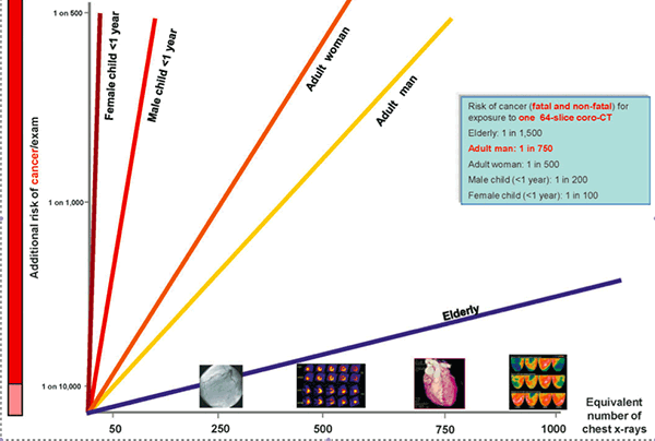
Figure 1. Risk of lifetime cancer risk and radiation exposure
Reproduced with permission from Malone J, Guleria R, Craven C, et al. Justification of diagnostic medical exposures: some practical issues. Report of an International Atomic Energy Agency Consultation. Br J Radiol 2012;85:523–38
It is also helpful to compare the risk of each test to everyday life. In Australia, the risk of dying from cancer before 85 years of age is 1 in 4 for males and 1 in 6 for females,9 and it is not possible to separate cancers occurring due to natural radiation, medical or other causes. The additional risk from 2-view chest X-ray examination for a male, aged 20 years, is 0.001%, or 1 in 90 000. In comparison, the lifetime risk of dying as a result of a motor vehicle incident in the United States is approximately 1 in 108.10
Table 3 relates the lifetime risk of cancer to lifetime risk of death from ‘everyday’ events. The additional risk of cancer, both fatal and non-fatal, posed by a single CT of the abdomen is similar to the risk of death from drowning.
Optimisation
Radiation protection is not just the responsibility of the primary care or referring doctor. Optimisation refers not only to technology, but also to auditing compliance with guidelines and avoiding unnecessary exposure by substituting other procedures.6,7 The Royal Australasian College of Radiologists and the Australian government have recently established diagnostic reference levels for CT, which are designed to help lower overall community exposure.5
Summary
Medical radiation has brought significant benefits to society. The risk of adverse effects, including cancer, from diagnostic radiology procedures is small, but the use of medical radiation, especially CT, is steadily rising. Before referring a patient for a medical imaging test that uses ionising radiation, the GP should ensure that the procedure is justified, and that alternative tests not using ionising radiation are inappropriate. General practitioners should be confident in communicating the relative risk of induction of cancer related to medical radiation with their patients, so that the patient can make an informed choice.
Useful resources
- Evidence based clinical decision support tool and educational resource for diagnostic imaging: www.imagingpathways.health.wa.gov.au
- Risk calculator for common medical imaging procedures with comparative risk tables: www.xrayrisk.com
- Health Physics Society – information specifically directed at physicians and patients: www.hps.org.
Competing interests: None.
Provenance and peer review: Commissioned; externally peer reviewed.