Classification of venous disease
Important changes have been made in the adoption of reporting standards in venous disease over the past few years, which will enhance our ability to gather data and compare outcomes across studies. The ‘basic’ Clinical impact, Etiology, Anatomy and Pathophysiology (CEAP) classification (Table 1), and other scoring systems are now well established tools for grading the severity of venous disease, as well as outcome measurement.2
Table 1. CEAP classification – C (clinical component only)2
| C0 |
No visible or palpable signs of venous disease |
| C1 |
Telangiectases or reticular veins |
| C2 |
Varicose veins |
| C3 |
Oedema |
| C4 |
Pigmentation, eczema, lipodermatosclerosis, atrophie blanche |
| C5 |
Healed venous ulcer |
| C6 |
Active venous ulcer |
| CA |
Asymptomatic |
| CS |
Symptomatic |
Multiple eponyms previously used to describe venous structures have been replaced with more intuitive, anatomically based names, and these have allowed for standardised abbreviations. Changes include:
- the confusing term ‘superficial femoral vein’ is now simply referred to as the ‘femoral vein’
- ‘short’ or ‘lesser’ saphenous vein is now referred to as the ‘small saphenous vein’ (SSV)
- ‘greater’ or ‘long’ saphenous vein is now referred to as the ‘great saphenous vein’ (GSV).2
Why do people get varicose veins?
Many causes of varicose veins are postulated, but only a genetic link and past history of deep vein thrombosis are supported by good evidence. The theory that the highest groin valve undergoes degeneration, and the rest then follow, makes good intuitive sense, but is not well supported by evidence: some 40% of patients with varicose veins do not have an incompetent sapheno-femoral junction. In some patients, there is a loss of elastic tissue in the vein wall, causing progressive incompetence of venous valves in the axial veins resulting in venous hypertension, reflux and total dilatation, causing varicosities.1
Symptoms
Common symptoms of varicose veins include:
- a cosmetically displeasing appearance – venous dilatation
- pain
- ankle swelling
- skin pigmentation
- eczema.
Less common symptoms include ulceration, superficial thrombophlebitis and haemorrhage.
Venous dilatation
Inframalleolar ankle flare or corona phlebectatica is the most common initial manifestation of venous disease (CEAP Class 1).
Isolated calf varicosities are commonly noted with prolonged standing or during menses. With progression of venous disease, the veins become more tortuous and distended, and patients may note their appearance in the proximal portion of the limb. Many women report that their varicosities progress rapidly in size and number during their first pregnancy (CEAP Class 2).
Oedema is an early symptom of venous disease (CEAP Class 3). This usually spares the metatarsal area. It has often been taught that only lymphoedema fails to pit on examination, however, non-pitting oedema is the result of subcutaneous fibrosis and repeated infections, irrespective of a venous or lymphatic cause.
Limb heaviness or ache that occurs after prolonged standing, and eases on walking or elevation, is typical of venous disease, whereas the claudication-type pain of arterial disease worsens on exertion. In severe chronic venous insufficiency with outflow obstruction, venous claudication may occur – in this situation the patient may describe a ‘bursting’ pain on walking.
Patients will commonly also experience pain and tenderness along the course of dilated varicosities. Patients with such severe reflux may also have severe skin pigmentation changes (CEAP Class 4).
Lipodermatosclerosis dramatically reduces the integrity of the affected skin, and minor trauma can result in ulcer formation – the end stage of chronic venous disease (CEAP Class 6).3
Physical examination
The physical examination should include inspection of the limb in a standing position. Notation should be made of:
- ankle venous flare
- telangiectasias (dilated interdermal venules <1 mm)
- reticular veins (non-palpable subdermal veins 1–3 mm)
- varicose veins (>3 mm)
- oedema
- skin pigmentation
- venous ulcers – chart on a diagram and note size, depth, character of the ulcer base.
Photo documentation is increasingly being used to:
- evaluate the response to therapy
- track the natural progression of the disease
- decide when to refer
- keep a medicolegal record (remember to get consent from the patient to take and store a photograph as part of the medical record).
It is also important to document arterial pulses in all patients. In unusually sited or appearing varicosities, consider anteriovenous malformation. Traditional non-invasive office based testing (eg. Trendelenberg) has largely been replaced by duplex ultrasound.
Diagnostic testing
Venous duplex ultrasound has become the standard of care for the investigation of varicose veins. Duplex ultrasound should document the:
- patency and competency of the deep system
- patency and competency of the superficial system, including site of incompetent segments, site of junctions and perforators
- location and relationships of the sapheno-popliteal junction
- diameter of the affected vein – this may be important in some endoluminal applications.
Decision making
Varicose veins are a progressive disease and will steadily worsen. Complications develop in a relatively small number of cases, and may prompt the patient to seek medical care. Many patients simply require some reassurance and explanation regarding the natural history of the disease.
Uncomplicated veins, without significant pain, can safely be managed with reassurance only. However, the ‘tea party’ system of referral is common, where patients have a relative or friend who has had a particular type of treatment, and have their mind set on having a similar treatment. It is also common for patients to have conducted their own web based research, and to have decided in advance which type of treatment they want. Although these patients may often not have a definite medical indication for intervention, it is often quite difficult to persuade them otherwise, and this may be a good reason to refer them for a specialist opinion.
There is extensive debate on the treatment of patients with uncomplicated varicose veins in the public health system, with much to be said for a very conservative approach in this category of patients. Some funding agencies will not support payment for this group. Many public guidelines, including Australian guidelines, suggest that only patients with advanced disease or symptoms should be treated in the public system (CEAP greater than 3).3
All patients with venous eczema or ulceration, evidence of chronic venous insufficiency, thrombophlebitis, bleeding, or severe discomfort, should be referred to a vascular team for assessment.4
A preliminary discussion with the patient before referral is often very useful, as some patients are resistant to any further intervention, and therefore it doesn’t make any sense to further investigate or refer on for anything other than compression therapy. As long as a patient has easily palpable foot pulses or an ankle-brachial index over 0.6, it is safe to fit Class 2 below-the-knee compression stockings. These will provide great relief for the symptoms of chronic venous insufficiency, and will control most varicose veins.
It is also important to consider the patient’s comorbidities. It is unlikely that patients with severe medical comorbidities or obesity will be offered surgical treatment, unless they have non-healing venous ulcers. This may change in the future with progression of the endovenous field.
Treatment options
Choice of treatment depends on many factors, including local expertise.5 Taking into account age, general health and symptomatology, patients with varicose veins may be offered one or more of the following management options:
- reassurance only
- conservative management with compression therapy
- endovenous therapy
- sclerotherapy – using liquid or foam (can be ultrasound guided, UGFS)
- endovenous laser therapy (EVLT)
- radiofrequency ablation (RFA)
- mechanical – using steam or rotating catheter
- surgical treatment.
It is difficult to make comparisons between the different treatment modalities, as there are few randomised controlled clinical trials. There is also a lack of standardisation of indications for surgery, intervention techniques, outcome measures, or long term data.
While open surgery remains the ‘gold standard’, most veins can be treated successfully with a range of options. There is a clear worldwide trend toward less invasive methods of treatment for varicose veins, with rates for laser, radiofrequency and sclerotherapy increasing every year. The recent addition of Medicare item numbers for laser and radiofrequency will most likely result in a similar trend in Australia.
A recent review of the literature revealed the following:
- no significant difference in short term outcomes between the modalities
- complications were few and mostly minor
- new varices may develop in 10% of patients within the first year after any treatment
- the technical failure rate was highest with UGFS
- both RFA and UGFS were associated with a faster recovery and less postoperative pain than EVLT and stripping
- the shortest time to return to work was seen in the UGFS and RFA groups
- there were significantly more cases of superficial phlebitis in the UGFS and RFA groups
- the mean cost per treatment was lowest in the UGFS group and highest in the RFA and EVLT groups.6–8
Recent guidelines recommend against UGFS alone for the treatment of GSV incompetence.3
In trying to advise the individual patient on how to proceed, it is helpful to consider which patients should not undergo endoluminal treatment. This would apply in cases where the varicose veins are:
- overly large (>2 cm diameter) – thermal treatment may fail, and there is a high risk of deep vein thrombosis
- overly tortuous – endoluminal catheters may not pass up the vein
- very close to the skin – skin burns are more likely, and in cases of thrombophlebitis – endoluminal catheters may not pass up the vein.
Economic considerations are also relevant. As with all new minimally invasive therapies, there will inevitably be trade, patient, media and operator driven pressure for increased uptake. It is incumbent on all of us to ensure that simply because the intervention is less invasive, its use is not extended to patients who have borderline or no indications for intervention.
Open surgery
Surgical interventions should be individualised according to the patient’s preoperative evaluation. An important principle is to ‘treat the patient and not the duplex scan’. A combination of ligation, axial stripping, and stab phlebectomy may be applied as needed to the GSV, SSV, tributary veins and perforating veins. Great saphenous vein stripping is commonly perceived as a painful and morbid procedure by patients and referring physicians alike. The memory of large incisions, extensive bruising, significant pain and prolonged disability from older techniques is of major concern to patients and referring physicians. However, recent refinements of technique allows for open surgery to be performed with the some of the advantages traditionally associated with the more minimally invasive techniques, including local anaesthesia/sedation with tumescent local anaesthesia, day case procedure, early mobilisation, minimal scarring, duplex ultrasound intra-operative assessment of completeness of surgery, and shorter duration of post-operative compression stockings.
In addition, recent studies have shown that gentle tissue handling in the groin incision results in less neovascularisation and less recurrence. With the advent of duplex mapping of incompetent veins preoperatively, the surgeon will have a much better idea of whether stripping of the GSV is necessary. In the 70% of patients who do have an incompetent thigh vein, high ligation alone will result in a very high recurrence rate, and is one of the reasons for the current poor press for open surgery.9 However, this should be viewed not as a failure of the technique, but as a failure of the operator.
Endovenous thermal ablation
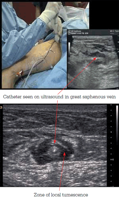
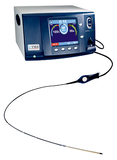
Laser and radiofrequency methods use similar techniques (Figures 1 and 2). Under sedation, and ultrasound guidance, large volume saline and local anaesthetic tumescence is created around the laser or RFA catheter, which is in the lumen of the GSV. A heat source (either laser at various wavelengths or radiofrequency) is then delivered through the catheter to the vein wall, with resultant coagulative necrosis. The saline tumescence creates a heat sink and protects the surrounding tissues from thermal damage.

Figure 1. Endovenous thermal ablation technique

Figure 2. Radiofrequency ablation machine
Injection sclerotherapy
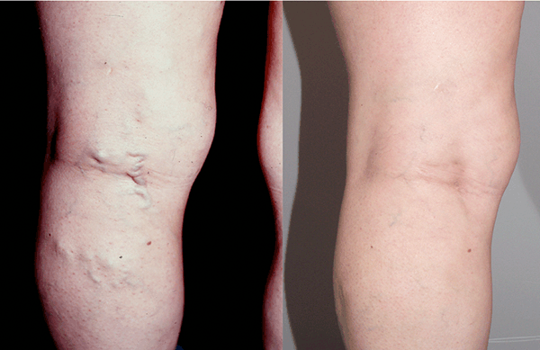
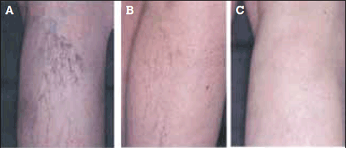
Numerous methods of injections and compounds are available. The most commonly used are sodium tetradecyl sulphate, polidocanol and aethoxysklerol (Figures 3 and 4). These can be foamed or injected in various concentrations. Foam can be prepared by hand or delivered from commercial canisters. A small amount of foam is usually injected at the sapheno-femoral junction under ultrasound guidance. This results in intense venospasm, subsequent contact with the vein wall, and sclerosis then occurs. Compression is applied and successive segments treated; 8 mL of foam is generally the maximum used.

Figure 3. CEAP 2 veins: Results of sclerotherapy – before treatment left, 1 year after treatment, right

Figure 4. CEAP 1 veins: Results of sclerotherapy – A) before treatment; B) 3 months after treatment; C) 1 year after treatment
Summary
Depending on age, general health condition, and symptomatology, patients with varicose veins may be offered one of a number of interventions. There have been important changes to venous practice in the past few years, and we need to choose our interventions carefully and monitor outcomes in order to ensure that patients get the most appropriate and cost effective care.
Key points
- Important changes have been adopted in the classification, scoring and anatomical notation of varicose veins in the past few years.
- A quality of life or degree of disability assessment is an important part of the initial consultation.
- Many patients simply require reassurance, and a thorough discussion of options at the primary care level may circumvent unnecessary referral.
- Compression stockings alone may be appropriate for patients who are too unfit for intervention or those who do not wish to have any form of surgical intervention.
- Many patients are treated for cosmetic concerns alone, so it is important to manage patients’ expectations.
- Minimally invasive treatment options such as injection sclerotherapy and endovenous modalities are becoming increasingly popular and have shown equivalence in short term outcomes.
- Conventional open surgery has also improved, with better outcomes, smaller incisions and duplex mapping.
- Patients with complications of varicose veins (CEAP 3–6); and those with clinical evidence of chronic deep vein insufficiency, especially venous eczema or ulceration, require referral to a vascular surgeon.
Competing interests: None.
Provenance and peer review: Commissioned; externally peer reviewed.
Acknowledgement
All images are courtesy of Mr Philip Coleridge Smith DM, FRCS, Reader in surgery and consultant vascular surgeon, British Vein Institute, London.