Case study
Bert, aged 80 years, presented to his general practitioner with sudden, painless loss of vision in his right eye 2 hours prior. He was an insulin-dependant type 2 diabetic, with a history of hypertension and dyslipidaemia.
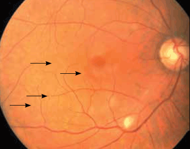
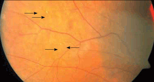
Right visual acuity consisted only of hand movements, compared to 6/6 vision in the left eye. There was a right relative afferent pupillary defect. Dilated fundus examination demonstrated multiple emboli in the arterial circulation, early arteriolar attenuation, and a cottonwool spot along the infero-temporal vascular arcade (Figure 1 and 2). Left fundus was normal.

Figure 1. Right fundus image of the posterior pole, demonstrating multiple retinal emboli (marked by arrowheads), an inferotemporal cottonwool spot and early arteriolar attenuation

Figure 2. Right fundus image of retinal periphery, demonstrating further multiple retinal emboli (marked by arrowheads)
Question 1
What is the differential diagnosis for sudden, painless, unilateral loss of vision in a patient of this age group?
Question 2
Given the presence of intra-arterial retinal emboli, what is the most likely diagnosis?
Question 3
What immediate investigations and management steps are appropriate?
Answer 1
The differential diagnosis for sudden, painless, unilateral vision loss in a patient of this age is:
- giant cell arteritis
- central or branch retinal artery occlusion
- nonarteritic ischaemic optic neuropathy
- retinal detachment
- central or branch retinal vein occlusion
- vitreous haemorrhage.
Answer 2
Given the presence of multiple intra-arterial retinal emboli along various vessels, the most likely diagnosis is an acute central retinal artery occlusion (CRAO). As a CRAO progresses, the arterioles appear attenuated and the retina becomes increasingly ischaemic. Eventually, retinal infarction and intracellular oedema cause retinal pallor. The macula appears deep orange-red in comparison to the surrounding pale retina – the classically described ‘cherry-red spot’. As this patient presented only 2 hours after onset, the retina had not yet become pale. Early recognition and intervention may be of benefit to the final visual outcome.
Answer 3
An urgent erythrocyte sedimentation rate (ESR) and C-reactive protein (CRP) should be performed to detect giant cell arteritis, but this should not delay referral to an opthalmologist. General practitioners in a rural setting may consider prescribing medications to lower the intraocular pressure after discussion with an ophthalmologist in cases where transfer may be lengthy or delayed. In such situations, digital ocular massage against a closed lid can also be administered by the patient or by the GP.1
Case study continued
Bert was referred immediately to a tertiary centre for urgent ophthalmology review. Ocular massage using a contact gonioscopy lens was performed. Intravenous acetazolamide and topical medications were given to lower the intraocular pressure. An anterior chamber paracentesis was performed, in which 0.1 mL of aqueous humour was removed using a 25 gauge needle via a limbal approach.
Bert also underwent urgent investigation to identify the source of the emboli. Cardiac echocardiography demonstrated no evidence of an intramural thrombus. However, carotid Doppler ultrasonography revealed a critical right internal carotid artery stenosis of 80–99% (Figure 3).
Bert was commenced on aspirin and intravenous heparin and underwent a right carotid endarterectomy 2 days later. Despite timely efforts to improve retinal perfusion, his right visual acuity did not improve.

Figure 3. Doppler ultrasound demonstrating a high grade stenosis (80–99%) of the right internal carotid artery (arrows)
Question 4
What are the causes of central retinal artery occlusion?
Question 5
What is the evidence for the management of central retinal artery occlusion?
Answer 4
Central retinal artery occlusion is an acute blockage of the central retinal artery that results in sudden, painless loss of vision. The central retinal artery is a branch of the ophthalmic artery and supplies the prelaminar optic nerve and the inner two-thirds of the retina.2
The most common cause of CRAO is thrombus or embolus from the carotid arteries or heart, which lodges in the central retinal artery. Risk factors are identical to those for atherosclerotic disease, and include diabetes mellitus, hypertension, hypercholesterolaemia and smoking.3
Vasculitides such as giant cell arteritis (which should be suspected in all patients over 50 years of age), systemic lupus erythematosis, Wegener granulomatosis and polyarteritis nodosa may also cause CRAO.
In younger patients, rare causes such as hypercoaguable states, sickle cell disease and paraneoplastic syndromes should be considered.4
Answer 5
While there is no proven treatment for CRAO, and visual prognosis is often poor, there are anecdotal reports for various strategies if employed within hours of the onset of symptoms.5 Most patients who have acutely and profoundly loss of vision are willing to undertake treatment that may be of some benefit. In our clinical experience, strategies to improve ocular perfusion by lowering the intraocular pressure can lead to visual improvement in some cases if performed within a few hours of onset of symptoms. Ocular massage, anterior chamber paracentesis and intraocular pressure lowering medications were tried in this case.
There have been reports of using intra-arterial thrombolysis with super-selective catheterisation of the ophthalmic artery to treat CRAO.6 However, there are concerns regarding haemorrhagic side effects and a recent small randomised control trial showed no benefit in visual acuity at 6 months compared to placebo.7 Other treatment modalities, including hyperbaric oxygen therapy8 and laser embolectomy,9 are based on small retrospective case series with varying degrees of efficacy.
Central retinal artery occlusion may be the first manifestation of atherosclerotic disease, preceding a larger cerebrovascular or cardiovascular event.4,10 One study found that 18.8%11 of patients with acute retinal artery occlusion had haemodynamically significant carotid artery disease. Investigation of arthymias and valvular disease through electrocardiographic monitoring and cardiac echocardiography are also needed.
Although there are no formal studies identified, evidence from the stroke literature would suggest that all patients who have sustained a CRAO in one eye should be on long term aspirin to reduce the risk in the other eye.5
Key points
- Central retinal artery occlusion is an important cause of sudden, unilateral, painless visual loss.
- Current treatments for CRAO involve time-critical attempts to reduce ocular pressure and improve ocular blood flow.
- Giant cell arteritis and embolic carotid disease are common causes of CRAO.
- Long term management includes regular aspirin and atherosclerotic risk factor modification.
Competing interests: None.
Provenance and peer review: Not commissioned; externally peer reviewed.