Case
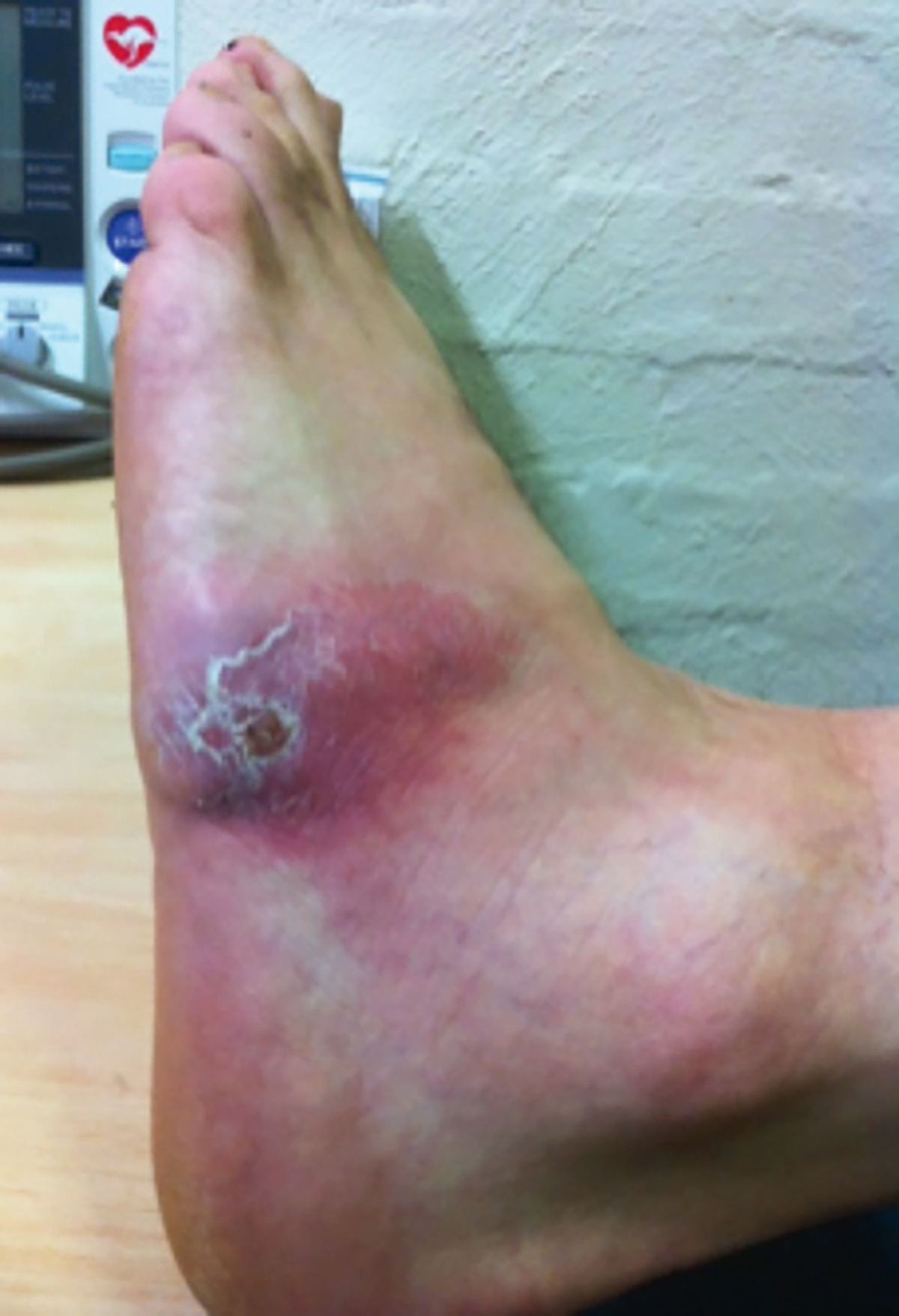
A man, 49 years of age, presented to a general practitioner (GP) with 10 days of worsening pain in his left foot. Six months earlier, he had sustained an injury from a garden rake that penetrated his foot (Figure 1). A GP who was consulted at the time of the initial injury provided tetanus immunisation and prescribed oral antibiotics.

Figure 1. Initial presentation
One month after the initial injury, the patient presented to another GP when he noticed that the scar site had become thickened and intermittently tender. A diagnosis of keloid formation was made and three corticosteroid injections were prescribed at intervals. These were noted to improve the appearance of the scar.
At this third presentation (six months after the initial injury), the patient was referred for an X-ray, which showed no signs of osteomyelitis. Blood tests, including full blood evaluation, C-reactive protein levels and erythrocyte sedimentation rate, were normal. An ultrasound (for embedded foreign body) showed inflammatory changes and a potential arteriovenous fistula formation, but was not suggestive of a specific pathology.
The patient had asthma, for which budesonide/formoterol had been prescribed, and was a current smoker. He was otherwise in good health.
Question 1
What is the most likely diagnosis and appropriate management?
Question 2
In what situations would wound biopsies be warranted? What is the role of corticosteroid injections?
Answer 1
The most likely diagnosis is a chronic wound infection and a search for the pathogen should ensue.
Answer 2
The first indication of atypical wound healing was the development of a keloid scar within one month of injury. The unresponsive nature of the wound should have warranted a biopsy at the second GP visit.
The Australian Therapeutic Guidelines (Guidelines) indicate the use of swabs if there is clinical suspicion of invasive infection requiring systemic therapy.1 The Guidelines recommend biopsy if the wound has an atypical appearance or location, or has not responded to therapy.1 A biopsy may be preferred in this case as swabs may be contaminated by surface-colonising organisms.2
Glucocorticoid injections are indicated for keloid and hypertrophic scars;3,4 however, they should only be prescribed if the clinician is confident that there is no infection.5 There should be a low threshold for the suspicion of an infection in a hypertrophic or keloid scar following a penetrating wound infection, as infection is thought to contribute to hypertrophic scar development.4
Case continued
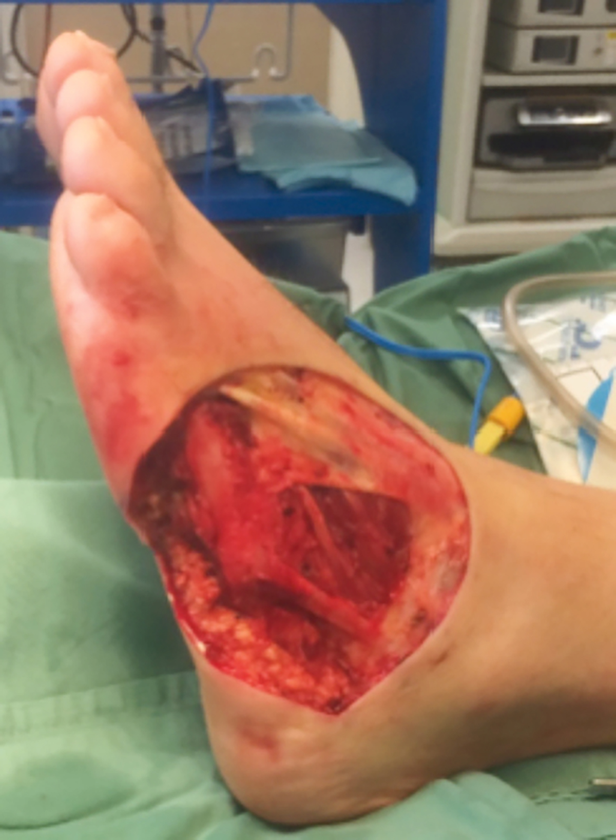
The patient was referred to a plastic surgeon and admitted to a private hospital for debridement and parenteral medications. Swabs taken during debridement tested positive for Scedosporium prolificans. Repeat debridement procedures were performed (Figure 2) and the final wound bed was covered with a temporary split-thickness skin graft. The patient was discharged on day 25 with parenteral antifungal treatment (voriconazole) and ongoing wound care. He subsequently received a myocutaneous flap and continues to do well.

Figure 2. Wound post-debridement
Question 3
The patient was referred to a plastic surgeon and admitted to a private hospital for debridement and parenteral medications. Swabs taken during debridement tested positive for Scedosporium prolificans. Repeat debridement procedures were performed (Figure 2) and the final wound bed was covered with a temporary split-thickness skin graft. The patient was discharged on day 25 with parenteral antifungal treatment (voriconazole) and ongoing wound care. He subsequently received a myocutaneous flap and continues to do well.
Answer 3
S. prolificans is a virulent fungal organism that can cause life-threatening infection in immunocompromised hosts. It is widely prevalent in the natural environment, particularly in soil and water. Although human infection is rare, its incidence is increasing, particularly in immunocompromised people.6 S. prolificans infection is usually contracted by inhalation of spores, or direct inoculation via a penetrating injury. The most common sites of infection are the respiratory tract, soft tissue, bones, joints, eyes and central nervous system (CNS).6,7 Progression to disseminated infection can rarely occur, particularly in immunocompromised patients, and may lead to CNS, pulmonary, skin and renal involvement.7 This patient’s presentation is not characteristic of mycetoma (eg swelling, sinus, fungal grain formation). This is typically a chronic, progressive and destructive infection that may spread to ligaments, cartilage and bone over time.6,8
There is no established standard treatment for S. prolificans because of antifungal resistance and the high rate of treatment failure.9 Therapy usually involves a combination of antifungal medication, surgical excision (where possible) and optimisation of the patient’s immune status.6,9 The prognosis for an individual patient is largely dependent on their immune status, the location of the infection and whether surgical debridement is possible.
Key points
- S. prolificans is a rare but virulent fungal organism that can cause life-threatening infection, particularly in immunocompromised hosts.
- Differential diagnoses other than common bacterial causes should be considered in chronic wound infections, particularly if unresponsive to empirical antibiotic therapy.
- Steroid injections should be given only if the clinician is confident that there is no infection.
- Wound biopsies are indicated if there is suspicion of infection, or if a wound is unresponsive or atypical in nature.
Authors
Brendan W Kite BSc, BComm (Hons), medical student, School of Medicine, Deakin University, Waurn Ponds, Vic
Terence Heng BAppSc, BCSc (Hons), MBBS, FRACGP, Vermont Health Care, Vermont, Vic. drterenceheng@gmail.com
Competing interests: None.
Provenance and peer review: Not commissioned, externally peer reviewed.