In community practice, patients who require further investigation have variable presentations but most commonly these include chest pain and/or dyspnoea. A chest X-ray (Figure 1) is a very useful initial study and allows exclusion of causes such as pneumothorax and pneumonia. Pulmonary and mediastinal masses may also be visible or suspected. Signs of cardiac failure may be present.
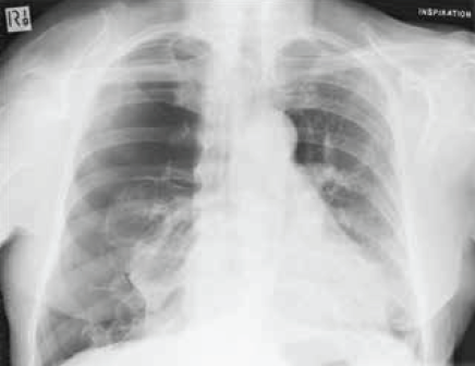
Figure 1. Chest X-ray. An X-ray can be diagnostic for causes of chest pain as in this image, which shows spontaneous pneumothorax
After exclusion of obvious non-cardiac causes for chest pain, myocardial perfusion study (MPS; Figure 2) is widely used and available for assessing whether reversible or fixed myocardial ischaemic changes are present. This study has a very high negative predictive score and allows stratification into three groups: obviously abnormal fixed or reversible ischaemia; normal; and equivocal, non- diagnostic.2 The group with equivocal findings are recommended for further evaluation with computed tomography coronary angiography (CTCA). The disadvantages of MPS include patient compliance with the exercise requirements of the study, radiation dose and relatively low sensitivity.
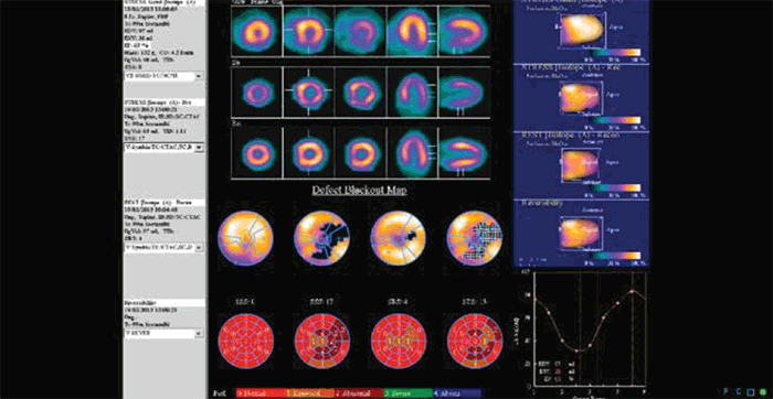
Figure 2. MPS of a patient presenting with atypical chest pain. The scan shows reversible ischaemia in the right coronary artery territory
Stress echocardiography is an alternative functional study that can be used for risk stratification purposes. This study does not require radiation exposure but the skilled and specialised technicians who perform this test are relatively ‘thin on the ground’ in some regions. Stress echocardiography does, however, provide details of wall motion/dysmotility that are not well demonstrated with other modalities. Stratification of results is similar to the MPS group.
CTCA
CTCA is conducted as an outpatient procedure and is non-invasive. The study achieves best results when the patient’s heart rate can be reduced to about 60 beats per minute. CTCA generally requires some premedication, usually with beta-blockers. An intravenous injection of non-ionic contrast is delivered via the cephalic or basilic vein in the cubital fossa. CTCA is generally well tolerated and does not cause complications, such as those related to arterial puncture, catheterisation and the passage of guidewires and catheters past the origin of carotid and cardiac vessels, which can occur with invasive or catheter angiography. The risks of iodinated contrast are the same as those of other intravenous contrast enhanced studies and are low.
The latest generation computed tomography (CT) scanners used for this examination require only one breathhold for the data acquisition component, which is usually completed in 8–15 seconds. The patient is then able to leave the scanning suite. Subsequent data and imaging manipulation is completed in 30–45 minutes. The radiation doses achieved with the latest scanners are of the order of only 1.5–3.5 mSv per study. These values compare favourably with normal yearly background radiation exposures at sea level and are comparable to or less than the levels experienced with invasive coronary angiography.
CTCA has a very high negative predictive value that approaches 100% in some studies and is thus very reliable in excluding CAD. It also has a lesser but very high sensitivity for demonstrating occlusive or high grade stenoses (Figure 3A, B).3
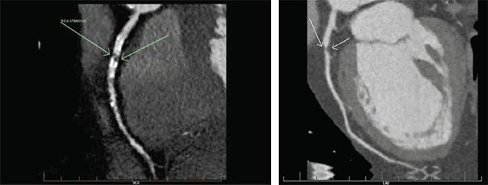
Figure 3. CTCA
A. High-grade stenosis is seen in the proximal right coronary artery (arrows). There is also extensive concurrent calcified and soft plaque elsewhere. A stent was inserted in this patient
B. 75% stenosis due to a soft plaque is seen in the left anterior descending coronary artery (arrows). The patient presented with atypical chest discomfort and was treated with an eluting stent
Limitations of the study include heavy coronary artery calcification, which obscures detail of the vessel lumen. Tachyarrhythmia also reduces the efficacy of the examination. In some centres, a preliminary CT calcium score study (non-contrast low-dose CT) is used to screen patients with too much calcified plaque to allow an adequate CTCA study. The coronary calcium score alone is also a useful tool in risk-stratification assessment in CAD.
CTCA also has the benefit of showing other cardiac and thoracic structures. The cardiac valves, chamber wall thickness and size, presence of filling defects, such as thrombi and tumour, can all be assessed in the same study. Mediastinal, hilar and adjacent lung lesions are also seen. The origin of coronary arteries can also be assessed; for example, the so-called ‘malignant origin’ of the anomalous right coronary arising between aorta and pulmonary artery is better diagnosed on CTCA than on any other study.
A variant of CTCA, known as the Triple Rule Out (TRO) study, is used in some centres in the USA to triage patients presenting with non-specific chest pain. This is commonly used in accident and emergency departments to exclude pulmonary embolus (PE), CAD and aortic dissection. TRO has high sensitivity and high negative predictive values for all three diagnoses but is not widely used at this time in the community setting in Australia.4 Currently, Medicare rebates for CTCA are available only when referred by a physician for defined indications and must be supervised and reported by an accredited specialist. The cost–benefit for this procedure has been evaluated overseas and there are data suggesting that the TRO can reduce hospital admissions and stays and obviate the need for other tests.5
Patients who have a past history of CAD and coronary artery bypass grafts (CABG) often pose a diagnostic challenge when presenting with chest pain. The patency of the coronary grafts and stents, however, is readily and quickly assessed with CTCA (Figure 4).
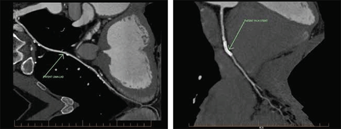
Figure 4. CTCA of grafts and stents
A. 2D angiogram showing LIMA–LAD graft patency (arrow). LIMA = left internal mammary artery; LAD = left anterior descending artery
B. CTCA showing patency of a stent inserted in the right coronary artery (arrow)
Discussion
Diagnosing significant ischaemic cardiac disease is a challenging and common clinical problem. In many instances, history and examination are sufficient to make the diagnosis, and simple tests, such as ECG and cardiac enzyme levels, can readily confirm the diagnosis.
A larger group of patients will require more investigation and imaging. The role of functional studies, including MPS and stress echocardiography, is well established and often allows a diagnosis of cardiac disease to be made or excluded. The technique of CTCA, recently introduced, has added another level of non-invasive diagnosis. CTCA, which has been refined and improved over the past 10–12 years, provides anatomical detail not previously available (ie. the presence of soft or non-calcified plaque). At the same time, CTCA assesses many other cardiac, mediastinal and thoracic structures. A diagnostic algorithm is shown in Figure 5. This algorithm is open to discussion, particularly if the early assessment is equivocal and if further development of CTCA, with its attendant benefit of full thorax evaluation, results in earlier usage. The future direction of CTCA technology will include greater speed, further reductions in radiation and contrast dose and more detailed plaque analysis.
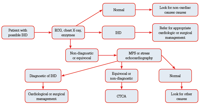
Figure 5. Diagnostic algorithm
The TRO study will become more available and probably cost-driven in acute triage situations. The role of MRI in coronary angiography is not comparable to CTCA at this stage, but current usage in chamber volume estimation, wall motion and perfusion is rapidly growing and in future may widely supplant other techniques that use radiation.
Summary
Diagnosing cardiac disease of ischaemic origin can be challenging in general practice. All patients do not, unfortunately, present with a clear-cut diagnosis. The role of imaging in identifying those with ischaemic heart disease (IHD) is an evolving process. This article has presented an initial functional or physiological approach with subsequent triaging of equivocal cases with CTCA. This approach may be supplanted in future by earlier CTCA imaging as this technique has the benefit of an inherent very high negative predictive score and a negative CTCA study can be immensely reassuring for our patients.
Competing interest: None.
Provenance and peer review: Commissioned; externally peer reviewed.