Case study
A man, 54 years of age, presented to our clinic with a 10 month history of bilateral interdigital lesions affecting the third and fourth interdigital spaces of both feet. Previous treatment had included multiple topical antifungal treatments and oral itraconazole, which had all been unsuccessful. He complained of a mild burning sensation in his toes.
Physical examination demonstrated malodorous interdigital lesions that on closer inspection were seen to be exudative erosions (Figure 1). There were no other relevant examination findings, and no relevant past medical history. The patient is a gardener who regularly wears boots.
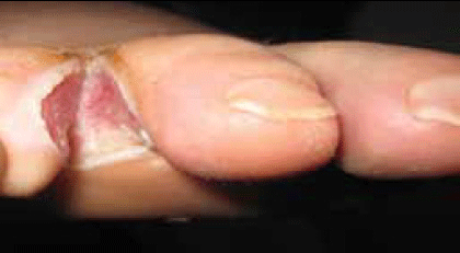
Figure 1. Third interdigital space of right foot with an erosive and exudative lesion on an erythematous background. Edges are white, due to maceration
Question 1
What is the diagnosis?
Question 2
What is the pathogenesis?
Question 3
What is the management of this clinical picture?
Question 4
What empirical treatment can be used for this condition?
Question 5
Are subsequent recommendations required?
Answer 1
Foot intertrigo: Bacterial interweb infection secondary to interdigital tinea pedis. In this case, the bacterial isolates were Pseudomonas aeruginosa and Trichophyton rubrum.
Gram negative bacteria are the most common causative agents among bacterial aetiology of this entity. P. aeruginosa is isolated most frequently. This is followed in frequency by Enterococcus coli and Proteus mirabilis. Gram positive cocci (Staphylococcus and Streptococcus) are less frequent agents.1
Answer 2
Multiple conditions can be involved in the pathogenesis of foot intertrigo. The interdigital space is colonised by polymicrobial flora, producing a precarious balanced cutaneous ecosystem.2 Damage to the stratum corneum of the skin from contact dermatitis, psoriasis, maceration, interdigital erythrasma or dermatophytosis can lead to a bacterial superinfection. Dermatophytosis also produces substances with antibiotic activity that can generate the selection of a resistant bacterial population.3
In the case presented, the lack of response to antifungal treatment along with malodour led us to initially suspect a bacterial aetiology. However, the location of lesions at the third and fourth interdigital spaces bilaterally is suggestive of an interdigital fungal infection. There were no pruritic symptoms reported, and no other lesions elsewhere on the body. In this context, we concluded the presence of dermatophytosis as a predisposing factor and a subsequent bacterial infection. This was confirmed by microbiological culture.
Less common causes of intertrigo include granular parakeratosis, bullous pemphigoid and Hailey-Hailey disease (a rare hereditary blistering skin disease).
Answer 3
Dermatophytosis, candidiasis and erythrasma can cause similar lesions. A history of persistent and repetitive treatment with topical and sometimes oral antifungals without clinical response may be suggestive of a nondermatophytic aetiology. Lesions in other intertriginous areas such as the groin or axilla can be seen in interdigital erythrasma. Dermatophytosis is suggested by bilateral involvement of the third and fourth interdigital space, lesions on the nails, and commonly in the inguinal crease (infection spread when the individual puts on his or her pants).
A Wood’s lamp is a useful screening test. Eythrasma generates a coral-red fluorescence (secondary to coproporphyrin III produced by Corynebacterium), whereas P. aeruginosa produces a green fluorescence (due to the pigment known as pyoverdin).4 Although the Wood’s lamp is useful in the examination of tinea capitis, is not useful in the case of dermatophytosis of glabrous skin. It should be noted that Wood’s lamp examination within a few hours of washing the area can generate false negatives as the substances that generate fluorescence will have been removed.
We recommend microbiological testing through potassium hydroxide mount of skin scrapings, gram stain or culture as clinical features have a low specificity. Ongoing follow up is necessary to evaluate the clinical response to treatment.
In the case presented, clinical response to oral ciprofloxacin (500 mg/twice daily) was satisfactory but dry intertriginous lesions persisted 15 days after initial treatment in the four interdigital spaces. These lesions were compatible with dry dermatophytosis lesions and microbial culture was positive for T. rubrum. Total resolution of the clinical picture was achieved through treatment with ciclopirox olamine cream.
Answer 4
Clinical manifestations of infectious foot intertrigo may not be specific enough for treatment.
If treatment is commenced before microbiological results are known, the following empirical regimen may be considered.
A localised infection may be treated with a topical antibacterial such as 5% amikacin; a widespread infection can be treated with oral agents such as flucloxacillin or ciprofloxacin.1
If the lesion is exudative, a povidone-iodine solution can be useful for its antiseptic and drying properties. A solution of 1:1000 or 1:10 000 potassium permagnate solution or Burrow’s solution can be used as a wet soak if blistering infections or discharging abscesses are present.
Answer 5
The patient should be appropriately educated to prevent recurrence. Patients with a history of dermatophytosis should increase the frequency of use of cleaning agents around the home, and consider wearing footwear in the shower recess. Attention to cleaning the feet and then drying them, especially the interdigital spaces, is recommended. A gentle antiperspirant or astringent may be beneficial in people prone to excessive sweating or who need to wear occlusive footwear. Patients with diabetic neuropathy should be encouraged to inspect their feet daily to identify any skin damage.
Conflict of interest: none declared