Case
A man, 74 years of age, presented with dactylitis on the first, second and fourth digits of his left hand, characterised by glossy erythema, oedema, pustules and yellowish scales. Additionally, there was onychodystrophy, subungual hyperkeratosis, almost complete onycholysis and pustules on the nail bed (Figure 1). The lesions first appeared two months ago and have been progressively worsening since, spreading proximally. There were no similar lesions and no evidence of psoriasis on full‑skin examination. The patient also noted functional incapacity of the affected digits, along with pain to the touch, but denied hand trauma prior to the appearance of the lesions.
His past medical history included arterial hypertension, type 2 diabetes mellitus, dyslipidaemia and chronic alcohol consumption. His medications included insulin glargine, amlodipine, acarbose and simvastatin.
Question 1
What differential diagnoses should be considered in this case?
Question 2
What is the most likely diagnosis in this patient?
Question 3
What is the aetiology of this dermatosis? How is it diagnosed?
Question 4
What are the available treatments for this condition?
Question 5
What is the prognosis of this skin disorder?
Answer 1
The main diagnoses to consider in this case are listed in Table 1.1–5
Answer 2
Acrodermatitis continua of Hallopeau (ACH), a localised pustular psoriasis, is the most likely diagnosis. It is a rare, chronic, relapsing condition, often triggered by local trauma or infection. ACH occurs in all age groups, but is more prevalent in middle-aged women.1,2
Answer 3
This dermatosis is considered a variant of pustular psoriasis; however, the aetiology is not completely understood. Inflammatory, infectious and neural aetiologies have been proposed. The diagnosis must be established on the basis of clinical features, and biopsy of the lesions may be performed. Other investigations to consider include Herpes simplex virus polymerase chain reaction (PCR) swab test, and Gram stain smear and culture of pustular fluid to exclude bacterial infection. A potassium hydroxide preparation of the pustules can exclude fungal infection.3
Answer 4
Because of the rarity of ACH and the limited evidence, mainly obtained from case reports, definite conclusions about the treatment are difficult to obtain, making the best approach unclear.6,7 Given the link to pustular psoriasis, many anti-psoriatic agents have been tried.3 Antibiotics and antifungal agents can be used if there is suspected infection. Usually, high or ultra-high potency topical corticosteroids are the first treatment choice, and can be applied once to twice daily over two to four weeks, preferably under occlusion. When adequate, the topical corticosteroid can be tapered to once or twice a week. Topical vitamin D analogues and calcineurin inhibitors can be used as adjunct or maintenance therapy, once or twice a week. If local treatment fails, topical psoralen and ultraviolet A (PUVA) therapy, narrowband UVB and systemic drugs (eg methotrexate, ciclosporin, acitretin) can be used. Although not approved by the Pharmaceutical Benefits Scheme, adalimumab, infliximab or etanercept may be attempted when there is no adequate response to any of the other treatments.3,6,8
Answer 5
ACH usually responds poorly to therapy, and spontaneous improvement is rare. There is no treatment that can ensure a long remission.1–7 In longstanding cases, osteolysis can occur, and generalised pustular psoriasis may develop, including von Zumbusch’s psoriasis, especially in the elderly.3,5
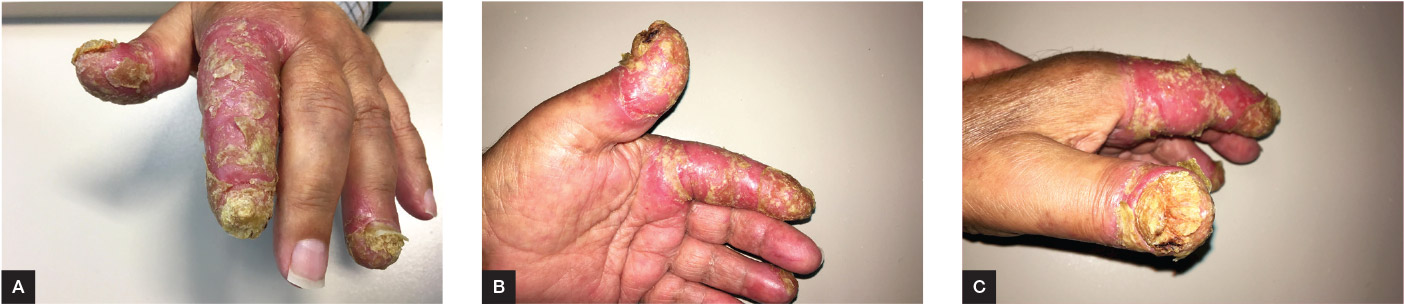
Figure 1. Clinical appearance of the lesions
A. Dorsal; B. Palmar; C. Lateral
Case continued
The skin biopsy revealed histopathological findings consistent with pustular psoriasis, which, along with the clinical presentation, supported the diagnosis of ACH. Laboratory tests found macrocytosis, anisocytosis, elevated transaminases, g-glutamyltransferase and hypercholesterolemia. Considering the comorbidities, laboratory abnormalities and extension of the lesions observed in the following appointment, combined treatment with betamethasone dipropionate 0.05% cream once daily, a cream containing ichthyol, salix alba extract, salicylic acid and provitamin D3 twice daily and topical PUVA was started.
The patient was re-evaluated three months later, showing no improvement of the disease. Therefore, treatment was changed to 0.05% clobetasol propionate under occlusion and 10% salicylic acid
in petrolatum ointment each applied once daily.
Table 1. Main differential diagnoses to be considered in the present case1–5
|
Disease
|
Clinical characteristics
|
|---|
|
Acrodermatitis continua of Hallopeau
|
Sterile pustules in an erythematous base, usually with desquamation
There may be onychodystrophy, anonychia or even osteolysis of the phalynx
More than one digit may be affected
Lesions start on their distal portion, spreading proximally over time
|
|
Staphylococcal infection
|
Usually as secondary skin infection of pre-existing lesions
Paronychia may be present
Small vesicles or pustules in an erythematous base
A yellowish crust may develop over the ruptured lesions
|
|
Herpetic whitlow
|
May exist a prodrome of fever several days before
Pain and edema of usually one digit
Vesicular lesions with clear fluid, clustered, in an erythematous base
There may be extension of the infection to the nail bed
|
|
Candidal paronychia
|
Acute paronychia is characterised by edema, erythema and tenderness of nail folds
Purulent material under the nail fold is common, which may drain if compressed. Onycodystrophy may occur
|
|
Pompholyx eczema
|
Itching and burning sensation of the hands and/or feet
Initially small vesicles with clear fluid may coalesce to form bullae
There is often subsequent desquamation
|
|
Dermatitis
|
Irritant dermatitis:
- Onset of symptoms: Minutes to weeks after exposure
- Itching, pain, macular erythema, scalded appearance and hyperkeratosis
Contact dermatitis:
- Onset of symptoms: Within days – longer if it’s the first contact
- Presentation change with the responsible agent
- Erythematous base with vesicles or papules
- Pruritic, hyperkeratotic plaques in chronic dermatitis
|
|
Squamous cell carcinoma
|
Mostly asymptomatic, the lesions may bleed or be painful
Macula, plaque or node with or without desquamation, hyperkeratosis and erosions
May be single or multiple lesions
|
|
Key points
- It is of the utmost importance that other diseases are excluded before making a firm diagnosis of ACH. Searching for infection is essential to the differential diagnosis, especially when there is no response to antibiotics.
- There is limited evidence regarding the treatment of this disease. Even when adequately treated, it is very difficult to achieve a lasting remission.
Authors
José Tiago Teixeira MD, Family Medicine Trainee, USF Viriato, Viseu, Portugal. jtiagopt@gmail.com
Carina Afonso MD, Family Medicine Trainee, USF Viriato, Viseu, Portugal
Maria Bernardete Machado MD, Family Medicine Trainee, USF Infante D. Henrique, Viseu, Portugal
Paulo Morais MD, Dermatologist, Department of Dermatovenereology, Centro Hospitalar Tondela-Viseu, Viseu, Portugal
Competing interests and funding: None.
Provenance and peer review: Not commissioned, externally peer reviewed.