Case study
A woman aged 59 years presented with a 12-month history of painful nodules on her fingers, which had recently worsened and become ulcerated. The patient had no dysphagia, dyspnoea, asthenia, muscle weakness or joint inflammation. Comorbidities included obesity, alcoholic liver disease, chronic renal failure, gastric antral vascular ectasia syndrome, anaemia, essential tremor and depression. Her regular medications included frusemide, lansoprazole, tiapride (a selective dopamine D2 and D3 receptor antagonist used to treat alcohol abuse, not available in Australia), ferrous sulfate, folic acid and sertraline.
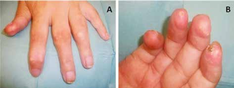
On examination, soft whitish-yellow papules and nodules were observed to affect mainly the finger pads and dorsum of the fingers bilaterally (Figure 1). Crusted ulceration was evident on the right index finger. There were no signs of Raynaud’s phenomenon, sclerodactyly, arthritis or other skin lesions.
 |
|
Figure 1. Clinical appearance of the lesions
|
|---|
A whitish fluid was extruded on punch biopsy, which was examined microscopically. Laboratory test results revealed normocytic normochromic anaemia, low albumin and renal dysfunction with urea 38.63 mmol/L and creatinine 0.23 mmol/L. Calcium levels were normal, but the uric acid level was 0.96 mmol/L.
Question 1
What is the most likely diagnosis in this patient?
Question 2
What is the aetiology of this condition?
Question 3
How is this condition diagnosed?
Question 4
What are the differential diagnoses?
Question 5
What is the appropriate management of this condition?
Answer 1
The most likely diagnosis is tophaceous gout. This condition is the result of monosodium urate crystal deposition in soft tissue, which can be the first sign of gout.
Answer 2
Gout is a metabolic disorder characterised by hyperuricaemia and deposition of monosodium urate crystals in the joints, kidneys and soft tissues, eventually resulting in arthritis, urate nephropathy and tophi.1,2 Some risk factors for gout include high purine consumption, alcohol intake, use of diuretics, cyclosporine and low-dose aspirin, obesity, hypertension and renal dysfunction.1–3 Without treatment, tophi classically appear about 10 years after the onset of the disease.4 However, they can develop without previous gouty arthritis.4,5
Answer 3
The clinical manifestations can be very suggestive, but they do not determine an accurate diagnosis of gout.5 Tophi present as papules or whitish-yellow nodules with a firm consistency. They are usually located on distal interphalangeal joints and tarsus, over the olecranon bursa and on the helix of the ear.2–6 Occasionally, they may ulcerate, releasing a lumpy whitish liquid5 and predisposing to secondary skin infection.
Tophi may be associated with destructive deforming arthritis. Nevertheless, radiographic findings are not specific for the diagnosis of gout.5
Serum uric acid levels are not enough for confirming or ruling out gout because they may be normal during an acute episode and there is a wide prevalence of asymptomatic hyperuricaemia.2,3
The most accurate way to diagnose gout is the demonstration of urate crystals in synovial fluid or in a tophus.2,5,7 However, a presumptive diagnosis of gout, on the basis of an accurate patient history and physical examination, can also be performed whenever the joint aspiration cannot be carried out.7
Answer 4
Table 1 shows conditions to be considered in the differential diagnosis of tophaceous gout. Other diagnoses that may be considered include ganglion cysts (digital myxoid cysts), pigmented villonodular synovitis, synovial chondromatosis and synovial sarcoma.4
Table 1. Conditions to consider in the differential diagnosis of tophaceous gout4–10
|
Disease
|
Differentiating features
|
|---|
| Rheumatoid nodules |
- History of rheumatoid arthritis
- On physical examination, rheumatoid nodules are frequently present on the extensor surface of joints and over pressure points
- Rheumatoid factor is usually positive
|
Calcium
pyrophosphate
deposition disease |
- Serum uric acid levels are normal
- Radiography findings include soft tissue swelling and chondrocalcinosis
- Microscopic analysis shows calcium pyrophosphate (CPP) crystals, which are smaller, rhomboid-shaped and have weakly positive birefringence
|
|
Calcinosis cutis
|
- May be the result of a variety of causes
- Histology shows granules and deposits of calcium in the dermis, often with a surrounding foreign-body giant cell reaction
|
|
Tuberous xanthomas
|
- They can be associated with dyslipidemias
- Nodules usually develop in pressure areas
- Histology reveals accumulation of vacuolated lipid-laden macrophages and multinucleated histiocytes
|
Answer 5
The patient should be advised on risk factor modification. Lifestyle measures include weight loss in obese patients, reduction in alcohol intake, avoidance of soft drinks sweetened with fructose, reduced consumption of purine-rich foods and adequate water intake.1,3 Consideration may be given to substitution of any medications associated with hyperuricaemia for alternative agents.
Pharmacotherapy is often necessary to prevent recurrence and chronic sequelae: urate-lowering pharmacotherapy is recommended for patients with more than two gouty attacks per year, in patients with tophi, urate nephropathy and in patients with joint damage seen on a radiograph.2,3 Allopurinol is the first-line urate-lowering therapy. Treatment should be initiated at 50 mg/day and increased gradually over 2–4 weeks, depending on the plasma urate levels. A dose of 300–600 mg/day may be required. Therapy should not be started during an acute attack. In patients with renal insufficiency, the allopurinol dose should be adjusted on the basis of the estimated glomerular filtration rate (eGFR). If eGFR is <30 mL/min the dose of allopurinol should be reduced to 100–200 mg/day; if eGFR is <10 mL/min, the dose should not exceed 100 mg/day.3 Concurrent treatment with low-dose colchicine (500 µg twice daily) or nonsteroidal anti-inflammatory drugs (NSAIDs) for the initial 3–6 months may reduce the risk of acute attacks.8,11 Alternatively, low-dose prednisolone can be used if other therapies are contraindicated.8 These agents are also used for symptom control during acute attacks of gout. Uricosuric agents are second-line therapy for allopurinol-intolerant patients or in cases of impaired renal excretion of urate.
Gout can be a debilitating disease leading to chronic pain, disability or chronic renal failure if not properly controlled.3 The importance of adherence to lifestyle modification and pharmacological therapy should be emphasised.
Case study follow-up
The histological study confirmed the diagnosis of tophaceous gout in our patient.She was advised to lose weight, and to reduce alcohol intake and consumption of purine-rich foods. The patient is now taking allopurinol 300 mg/day.
Competing interests: None.
Provenance and peer review: Not commissioned, externally peer reviewed.