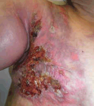
Figure 1. Clinical photograph of right chest wall 2 months after onset of symptoms. Note - this was taken 1 month after presentation to GP and Emergency Department

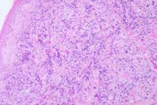
Figure 2. Haematoxylin and eosin stain of right chest wall punch biopsy showing invasion of dermis with cells consistent with invasive ductal carcinoma

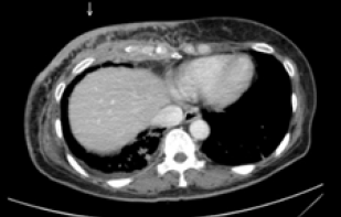
Figure 3. Computed tomography demonstrating significant enhancement of soft tissue throughout the right anterior chest wall involving cutaneous, subcutaneous and deep muscular tissues
Question 1
What is CEC?
Question 2
What is the management of CEC?
Question 3
What is the prognosis of CEC?
Question 4
What percentage of breast cancers have cutaneous manifestations?
Question 5
What are the different cutaneous manifestations of breast cancer?
Question 6
What are the most common primary malignancies in patients with skin metastases?
Answer 1
CEC is a rare form of metastatic cutaneous malignancy that is most commonly seen as a recurrence of breast cancer although it can present as a primary untreated breast cancer.1-3 It was first described in 1838 by Velpeau, as a stiff, leathery shield-like plaque due to its resemblance to the metal breast-plate of a cavalry soldier or cuirassier.2,4,5
It is characterised by diffuse morphea-like induration with scattered, lenticular papulo-nodules, overlying red-blue colouration and erythematous smooth cutaneous surfaces.4 Sclerodermatous plaques are formed with no associated inflammatory change.4 CEC involves a rapid dermal spread as it invades the cutaneous lymphatics and often involves large areas of chest wall, abdomen or, less commonly, the groin.3 It usually presents a few months or years after the primary carcinoma has been diagnosed and can appear clinically like an infectious cellulitis.1 Symptoms include pruritus, foul smelling discharge, oedema, bleeding and pain. The burning pain is likely to be caused by perineural lymphatic metastatic dissemination.5
Histologically, fibrosis is prominent with some cells displaying an ‘Indian file’ pattern where small lines between collagen bundles are formed by the tumour cells. It should be noted than an Indian file pattern is typical of invasive lobular carcinoma where as this case presented with a manifestation of invasive ductal carcinoma.6 The cells in CEC often appear similar to fibroblasts although they have larger, angulated and more deeply basophilic nuclei.2,4 There is decreased vascularity throughout, as the cancer disseminates along the tissue spaces through lymphatic vessels.3,5 The macroscopic induration is postulated to be caused by chronic lymphatic obstruction due to tumour emboli.5 In non-melanomatous cutaneous metastasis a punch or excisional biopsy is preferred to a superficial shave biopsy.7
Answer 2
Cutaneous metastases are often representative of extensive underlying disease, thus the principal aim of management of CEC is to improve the patient’s quality of life.3 Various methods, including intra-lesional chemotherapy, intra-venous chemotherapy, radiotherapy, skin grafts and hormonal antagonists, have been used to treat this illness with limited effectiveness.2,3 Theoretically, the extensive reactive fibrosis seen in the disease renders chemotherapeutic agents ineffective by preventing them from attaining tumoricidal concentrations locally.3 Prostaglandin synthetase inhibitors have also been used to decrease pruritus and pain by suppressing the prostaglandin-like material formed by the tumour.4 One promising study showed that pulsed brachytherapy resulted in local control of dermatologically metastasized breast cancer in 89% of patients.8
Answer 3
The prognosis of CEC depends on the type and biological behaviour of the underlying primary tumour, although skin metastases are usually associated with advanced disease.1 Generally, cutaneous metastases herald a poor prognosis. The average survival time of patients with skin metastases from any primary malignancy is approximately 7.5 months.9
Answer 4
Breast carcinoma is the most common cancer in women.4 In the largest reported series by Lookingbill et al., the incidence of cutaneous involvement in patients with breast carcinoma was 23.9%.9,10 Cutaneous metastases of breast carcinoma are the most common metastases seen by dermatologists.9
Answer 5
The most common cutaneous manifestations of breast carcinoma include nodular carcinoma (46.8%), alopecia neoplastica (12.0%), telangiectatic carcinoma (8.0%), malignant melanoma-like metastases (6.3%), carcinoma erysipelatoides (6.3%), subungual metastases (4.6%), CEC (4%) and zosteriform metastases (3.6%).11
Answer 6
Refer to Table 1. Cutaneous metastases are the first sign of internal malignancy in 37% of men and 6% of women.9
Table 1: Distribution of primary malignancies in patients with
skin metastases9
| Male | % | Female | % |
|---|
| Melanoma |
32.3 |
Breast |
70.7 |
| Lung |
11.8 |
Melanoma |
12.0 |
| Large Intestine |
11.0 |
Ovary |
3.3 |
| Oral Cavity |
8.7 |
Oral Cavity |
2.3 |
| Kidney |
4.7 |
Lung |
2.0 |
| Breast |
2.4 |
Large Intestine |
1.3 |
| Oesaphagus |
2.4 |
Endometrium |
1.3 |
| Other |
26.7 |
Other |
7.1 |
Key points
- It is important to consider skin metastases when assessing atypical dermatological findings in a patient with a history of cancer.
- Clinicians should be aware that cutaneous metastases from breast cancer can sometimes mimic the appearance of cellulitis.
Competing interests: None.
Provenance and peer review: Not commissioned; externally peer reviewed.