Table 1. Differential diagnosis of hip pain in young adults
| Extra-articular | Intra-articular |
|---|
Muscles
- Abductor muscle injuries
- Gluteus muscle tears
Nerves
- Sciatica
- Obturator nerve irritation
- LFCN irritation
- Piriformis syndrome
Tendons
- Snapping hip (ITB or iliopoas)
- Bursa
- Trochanteric bursitis
Ligaments
- Inguinal ligament strain
- Joint capsule*
Referred pain*
- Lumbar spine
- Knee
- Non-musculoskeletal pathology
|
Bones
- FAI
- OA*
- AVN*
- DDH*
- Fractures*
- Perthe’s*
- Septic arthritis*
Soft tissues
- Labral tear
- Chondral defect
- Ligamentum teres injury
|
ITB = iliotibial band, LFCN = lateral femoral cutaneous nerve, FAI = femoroacetabular impingement OA = osteoarthritis, AVN = avascular necrosis, DDH = developmental dysplasia of the hip.
*Not covered in this review |
Assessment
Patients typically present when their hip pain impairs activities such as work, exercise or sport. Symptoms suggestive of hip pathology include localised symptoms (such as catching sensations), symptoms related to activity or when going up and down stairs, or symptoms related to prolonged sitting or standing.
History helps to localise the hip as the source of pain rather than make a specific diagnosis as there is significant overlap in symptoms originating from different structures in and around the hip. For example, pain and tenderness over the greater trochanter, buttock or lateral thigh can suggest trochanteric bursitis, a tear of the gluteus medius muscle or a snapping hip.3 Patients with FAI most commonly report groin (88%), lateral hip (67%) and anterior thigh (35%) pain but may also complain of buttock (29%), knee (27%) and lower back (23%) pain.4 Other conditions that present predominantly with groin pain (eg. osteitis pubis, incipient inguinal hernia, adductor tendinopathies) have been the focus of a previous review article5 and are not addressed here.
Hip examination
The aim of a focused hip examination is to confirm the hip as the source of symptoms and to exclude alternative diagnoses such as referred pain rather than make a definitive diagnosis. Clinical examination has been shown to have a high sensitivity (98%) in localising intra-articular hip pathology but is poor in exactly defining its nature.6
Look
Inspection of the patient’s standing posture and gait will reveal any obvious asymmetry in the musculature or alignment. An antalgic gait (short stance phase relative to swing phase) reflects pain on weight bearing and may indicate a painful joint. The Trendelenberg gait reflects the integrity of the hip abductor muscles on the side of the standing leg. The patient can also indicate the site of symptoms.
The patient may have one of the clinical signs suggestive of intra-articular hip pathology (Figure 1). Cupping of the greater trochanter in the trochanteric C-sign7 (Figure 1A), pointing with two fingers towards the hip joint in the triangulation sign (Figure 1B) or pointing deep within the groin crease in the deep pointer sign (Figure 1C. It is important to note that these signs are commonly reported anecdotally rather than being evidence-based and their sensitivity for detecting intra-articular hip pathology is not known.
Figure 1. Clinical signs often performed by patients with FAI syndrome A) trochanteric C-sign, B) triangulation sign, C) deep pointer sign
Feel
Palpation may reproduce symptoms over anatomical landmarks suggestive of extra-articular pain. Pain reproduced by palpation over the greater trochanter is suggestive of trochanteric bursitis or a snapping hip (iliotibial band irritation over the greater trochanter). Buttock tenderness to palpation suggests muscular pathology (such as gluteus medius tear) and tenderness over the psoas tendon (located lateral to the femoral nerve just below the inguinal ligament) is suggestive of psoas tendonitis.
Move
Active range of movement (ROM) will test muscle integrity. Further assessment of specific muscle groups should be made where weakness or pain is identified. Passive ROM assesses the integrity of the joint and surrounding soft tissues, a reduction in which suggests FAI or labral/chondral injury in this patient group.
Special tests
Special tests are indicated where intra-articular hip pathology is suspected after exclusion of acute conditions that require emergency department referral (eg. septic arthritis, fracture, slipped upper femoral epiphysis (SUFE), dislocation). The anterior impingement test (flexion, adduction and internal rotation; Figure 2) and the FABER test (flexion, abduction and external rotation; Figure 3) have the highest sensitivities and specificities of the special tests available (>0.9) for detecting intra-articular hip pathology.8 A reproduction of symptoms, pain and a decreased ROM relative to the unaffected side represent a positive test result. Although >90% of patients with FAI will have a positive anterior impingement and FABER test, a positive test can indicate intra-articular hip pathology unrelated to FAI (eg. traumatic labral tears). These tests are therefore not diagnostic but aid in identifying intra-articular hip pathology.
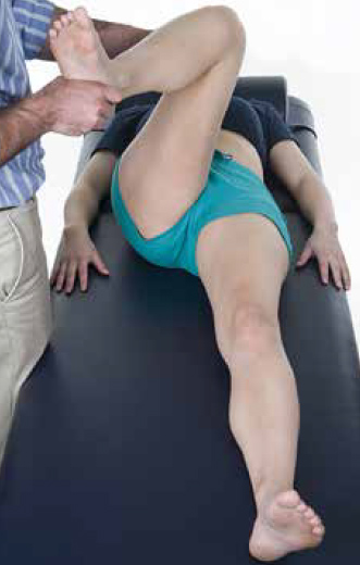
Figure 2. The anterior hip impingement test. The hip is positioned in flexion, adduction and internal rotation
Figure 3. The FABER test (flexion, abduction and external rotation)
Basic imaging
An anterior–posterior (AP) radiograph of the pelvis is an essential initial investigation to exclude fractures, developmental dysplasia of the hip, avascular necrosis, OA, malignancy and a missed childhood SUFE. However, for most conditions described in this review, the radiograph may be normal or show only subtle abnormalities that are easily overlooked.9 Figure 4 shows an AP pelvis in a patient with cam-type FAI. We suggest that additional views such as a Dunn view or frog leg lateral, and CT and MRI are best conducted by a specialist.
Figure 4. AP pelvis radiograph showing cam-type hip shape in FAI (left hip, red arrow)
Hip conditions in young adults
There are a number of conditions that may present with hip pain. Trochanteric bursitis typically presents with tenderness over the greater trochanter. The snapping hip originates from either the iliopsoas tendon or the iliotibial band (ITB). Snapping from the iliopsoas tendon is often audible and recreated when the hip is passively moved from flexion, abduction and external rotation to a position of extension with internal rotation.7 Snapping from the ITB is more visible than it is audible and patients often refer to the sensation of subluxation or dislocation as the tensor fascia lata ‘snaps’ back and forth across the greater trochanter.7 Gluteus muscle tears typically present with buttock pain, which is reproduced by palpation, but symptoms may also include pain over the greater trochanter and later hip pain. These pathologies can be grouped as the greater trochanteric pain syndrome in recognition that symptoms often overlap and are sometimes linked (eg. trochanteric bursitis due to a snapping hip).3 Patients typically present with pain and tenderness over the greater trochanter, buttock or lateral thigh. The prevalence is 1.8 per 1000 in the general population10 and although the incidence is thought to be low among young adults, this diagnosis should be considered where the symptoms are activity-related or follow injury.
Neuropathies causing symptoms around the hip joint include irritation of the sciatic nerve, obturator nerve and lateral femoral cutaneous nerve (LFCN) of the thigh. Symptoms include shooting pains, stinging or numbness, and neuropathic pain in the nerve distribution. They typically arise from nerve entrapment, such as piriformis syndrome and inguinal ligament strain, causing entrapment of the LFCN of the thigh.
The acetabular labrum is a cartilaginous ring surrounding the acetabulum and its function is to increase hip joint stability.11 Labral tears can arise from FAI, trauma, dysplasia, capsular laxity and degeneration.12 The ligamentum teres arises from the transverse ligament of the acetabulum and inserts into the fovea capitis of the femoral head. It is thought to provide stability, vascularity, proprioception, and nociception to the hip joint and ligamentum teres injury is recognised as a source of pain from the hip.13 Chondral defects refer to damage of the mature articular cartilage, which causes pain and may initiate the degenerative process of OA.14
FAI
The term FAI describes subtle deformities in hip shape that cause impingement between the femoral neck and anterior rim of the acetabulum during the normal range of functional hip movement, particularly in flexion adduction and internal rotation. The impinging surfaces can irritate and damage the soft tissues of the hip joint of which most at risk are the acetabular labrum and the adjacent acetabular cartilage. Hip shape deformities are classified into three types:15
- cam type – asphericity of the femoral head; widening of the femoral neck. The term comes from the cam-lobes on engine cam-shafts, which open and close valves by impinging on the appropriate surface as they rotate
- pincer type – over coverage of the anterosuperior acetabular wall; a deep socket. Similar to the tips of pincer forceps
- mixed type – a combination of cam and pincer deformities.
Cam impingement is more common in young men, and pincer in middle-aged women. Other types exist and are related to the orientation of the acetabulum and femoral neck. We use the term FAI syndrome to refer to patients with hip shape abnormalities and symptoms suggestive of impingement.
What is the prevalence of hip shape abnormality and FAI syndrome?
Hip shape abnormalities characteristic of FAI are quite common in the young adult population.16 In a prospective study of 200 asymptomatic volunteers aged 21–50 years, the prevalence of cam-type hip shape was found to be 14%.17 The prevalence of hip shape abnormality is reported to be higher in asymptomatic athletes than in the general population and the reasons for this remain unclear. A prospective study of American college football players (average age 21 years) found that 95% of the 134 asymptomatic hips had at least one radiological sign of cam or pincer shape18 and in a retrospective review of elite soccer players, radiographic hip abnormality was present in 72% of men and 50% of women.19 In patients with hip pain the prevalence of shape abnormality is even higher. A retrospective review of the pelvic radiographs of 157 patients aged 18–50 years revealed that 87% were found to have a hip shape abnormality.9
Why do some people get symptoms and others do not?
It is not yet understood why some people develop symptoms (FAI syndrome) and others do not. It is likely that the mechanism involves a combination of factors: a hip shape abnormality together with a level and type of activity that provokes impingement. There may also be a genetic predisposition to shape abnormality and/or soft tissue damage in these patients.20 The natural history of FAI and long-term progression to OA remain topics of much debate and ongoing research.
Management in primary care
Many conditions described in this review require diagnosis by a specialist. Many of these conditions respond to a course of non-operative care, particularly physical therapy, and there is no evidence that such treatment is harmful.21 Therefore, for young adults with persistent hip pain it would be reasonable to commence a course of physical therapy pending a diagnosis. In many cases the specialist will continue to treat non-arthritic hip pain with physical therapy, as failure to respond to this may well then be used as an indication to expedite surgery.22
For non-operative care, exercise-based physical therapy probably has the most evidence for effect but it is also reasonable to consider a short course of non steroidal anti-inflammatory drugs (NSAIDs), activity modification, education and advice, although limited evidence exists for this.21,23 Although non-arthritic hip pain is not a life- or limb-threatening condition, some causes, particularly FAI, are associated with an increased risk of OA. Therefore, it is advisable to obtain a specialist referral/diagnosis in a timely manner and within 3–6 months if symptoms do not improve with conservative management.
Early specialist referral may be indicated in athletes where the prevalence of hip shape abnormality has been shown to be substantially higher than in the general population.18,19 At present there is no evidence that patients with asymptomatic incidental findings of FAI benefit from any intervention, but patient education regarding presenting early if symptoms develop is advised.
Specialist assessment
Patients who attend a specialist will have a reassessment of their symptoms and clinical examination. They may also be asked to complete a validated hip score questionnaire to quantify their symptoms and monitor changes over time and treatment response. The specialist may use more detailed imaging techniques, such as magnetic resonance arthrography (MRA) and 3-dimensional CT, to diagnose soft tissue and bony pathology and to plan treatment.
When the diagnosis remains unclear or when multiple pathologies are suspected, a diagnostic intra-articular injection of local anaesthetic may be used. This has been shown to be an indicator of intra-articular pathology with an accuracy of 90%.6 Three-dimensional surface reconstructed CT provides the best impression of all aspects of hip shape and is particularly useful in pre-operative planning for FAI surgery (Figure 5).24
Figure 5. 3-Dimensional reconstructed CT images of cam-type deformity (red arrows)
Treatments often involve targeted physiotherapy, which has shown good short-term outcomes in pain and function for patients with mild FAI, although there is limited experimental data.21,25 The therapeutic aims are to increase the pain-free passive range of movement, improve the precision of hip motion, avoid hip hyperextension and femoroacetabular rotation under load, and to optimise the balance of muscle strength and length at the pelvis.25
Surgical management may be considered for extra- and intra-articular hip pathologies when patients do not improve with non-operative care and where the symptoms are judged severe enough to justify the risks of surgery. Trochanteric bursitis, the snapping hip, and focal isolated gluteus medius and minimis tendon tears can be treated effectively with arthroscopic surgery.26,27
Shape-corrective surgery for the treatment of FAI, as well as soft tissue repairs (eg. labral repair/reconstruction, microfracture and repair of ligamentum teres injuries) can be also be carried out arthroscopically.12,14,28 A growing body of literature now exists showing favourable short-to-mid-term outcomes of arthroscopic surgery for FAI in young adult and adolescent populations, although long-term data are still awaited and guidelines suggest that such surgery should only be carried out by specialists with expertise in arthroscopic hip surgery.29
Key points
- Persistent hip pain in young adults should not be ignored.
- Clinical examination and basic imaging are important to exclude conditions such as childhood hip disorders, OA, septic arthritis and fractures.
- Commence conservative management (NSAIDs, activity modification and physiotherapy) and follow up within 3 months.
- Refer patients with persistent hip pain of 3–6 months duration for specialist review and further investigation.
Competing interests: John O’Donnell has received payment for Board membership from Smith and Nephew. He has also received payment for consultancy from Arthrocare and Medacta, has grants pending from Arthrocare and has received royalties from Medacta.
Provenance and peer review: Not commissioned; externally peer reviewed.
Acknowledgements
The authors would like to thank the Medical Photography and Illustration Department at University Hospitals Coventry and Warwickshire NHS Trust for the production of images included in this review.
Patient consent was obtained for the publication of photographs in this review.