Neonatal skin is usually soft and smooth, covered with vernix caseosa, a derivative of sebaceous secretion and decomposing epidermal cells. Desquamation is usually delayed, occurring between 24 hours and 3 weeks, earlier in term than premature babies. Desquamation on day one may represent intrauterine anoxia or a disorder of keratinisation (ichthyosis). Colour usually rapidly becomes pink apart from the hands, feet and lips, where an initial dusky acrocyanosis may occur as a result of increased vascular tone.
Trauma at birth may give rise to a boggy scalp mass from oedema and venous congestion (caput succadaneum) or a subperiosteal haematoma (cephalohematoma), the latter limited to one cranial bone and possibly associated with an underlying fracture, secondary hyperbilirubinaemia, calcification and infection. Iatrogenic trauma can also cause scarring (eg. pits, dimples), anetoderma (localised loss of elastic tissue presenting as a soft protuberance), bleeding, infection, calcified nodules and aplasia cutis (absence of skin). Other causes of aplasia cutis are listed in Table 1, and when associated with glistening 'membranous' changes or a 'hair collar', there may be other neural tube defects.3 Children with aplasia cutis, where there is localised absence of intact skin at birth, should be referred for specialist assessment.
Table 1. Causes of aplasia cutis
| Genetic |
- Epidermolysis bullosa
- Syndromes: eg. chromosomal anomalies (Patau, Wolf-Hirshhorn), Delleman, Finlay-Mark, Goltz, Johnson-Blizzard, Kabuki, Setleis
|
| Developmental |
- Naevi (epidermal)
- Neurological malformations (eg. encephalocoele or myelomeningocoele)
- Adams-Oliver syndrome with limb anomalies
|
| Antenatal/perinatal event |
- Papyraceous fetus or placental infarct
- Trauma
|
| Infection |
- Toxoplasmosis, rubella, cytomegalovirus, herpes
|
| Drugs |
|
Neonatal colour
Harlequin colour change, presenting as reddening of the lower half and pallor of the upper half of a baby lying on the side, lasts for several minutes. It presents in the first 3 weeks of life, usually days 2–5, as a result of hypothalamic immaturity, and is more common in prematurity and intracranial injury.
Reticulated (livedoid) purplish mottling of the skin may represent physiological capillary and venular dilatation accentuated by cold (cutis mamorata), but may persist pathologically (Trisomies 18 and 21, Cornelia de Lange). Cutis mamorata telangiectatica congenita is a fixed vascular change associated with subtle dermal atrophy and sometimes, limb length discrepancy, which can be part of a syndrome. It may lighten with vascular laser treatment.
Neonatal birthmarks can be easily misdiagnosed: hemangiomas can resemble port wine stains, while early congenital melanocytic naevi may resemble café au lait macules. Appropriate referral and follow up is critical as the window of opportunity to treat haemangiomas (eg. with beta blockers) and giant congenital melanocytic naevi (with early curettage) may be very narrow.4,5 Melanocytic naevi that are medium to large or involve cosmetically sensitive areas should be referred for specialist management. For more details on birthmarks see the article by Ryan and Warren in this issue of Australian Family Physician. Other causes of colour change in the skin are listed in Table 2.
Table 2. Causes of neonatal skin discoloration
| Colour |
Age |
Causes |
| Jaundice |
|
Haemolysis (eg. ABO, rhesus), infection, drugs
Breastfeeding, cholestasis, enzyme deficiencies, sepsis, hepatitis, drugs, hypothyroidism, galactosemia |
| Bronze baby syndrome (grey-brown colour) |
First week |
Phototherapy photoproducts |
| Grey baby syndrome |
|
Chloramphenicol |
| Carbon baby syndrome |
|
Universal melanosis |
| Cyanosis |
|
Cardiorespiratory disease |
| Pallor |
|
Shock, asphyxia, anaemia, oedema |
Vesicular and pustular eruptions
Although the most common neonatal vesiculo-pustular eruptions are benign and self limiting, possible serious causes must be excluded, especially bacterial, viral and fungal infections (Table 3 and 4, Figure 1).6
Table 3. Causes of pustular neonatal eruptions
| Cause |
Age |
Investigations |
| Infectious |
- Bacterial: Staphylococcus aureus, Streptococcus pyogenes, Hemophilus influenzae, Escherichia coli, Klebsiella pneumoniae, pseudomonas, listeria
|
|
Swab MCS, systemic work-up |
- Syphilis (palmoplantar changes)
|
|
Darkfield microscopy serology, X-ray |
- Viral: herpes simplex virus, herpes varicella-zoster cytomegalovirus, AIDS
- Fungal: candida, pityrosporum
- Parasitic: scabies
|
First 6 weeks |
Tzanck/IF/PCR/ culture, urine sediment, serology
Swab MCS |
| Reactive |
|
|
First weeks |
Smear for stains
|
- Transient neonatal pustular melanosis
|
Day 0 |
|
|
|
Day 1–3 |
|
- Eosinophilic folliculitis
|
First year |
|
|
|
First year |
|
|
|
Hours to 6 weeks |
- neutrophils (+/– eosinophils
|
| Infiltrate |
- Histiocytosis
- Incontinentia pigmenti
|
|
Histology
- histiocytes
- eosinophilic spongiosis
|
| MCS = microscopy, culture, sensitivity; IF = immunofluorescence; PCR = polymerase chain reaction |
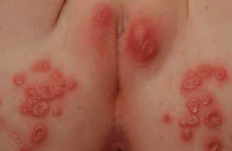
Figure 1. Clustered vesicles and uniformly small punched-out ulcers, herpes simplex
Erythema toxicum occurs more often in term babies of multigravidas. It usually arises in the first 4 days of life and fades within 4 days. Occasionally onset is delayed until 10 days after birth or it recurs in the first 2–6 weeks after birth. It is characterised by red macules and papules, with pustules appearing in a third of cases. Often widespread, it resembles 'flea bites', but spares the palms of the hands and soles of the feet. Folliculocentric subcorneal and intraepidermal pustules contain eosinophils and neutrophils (Gram/Wright/Giemsa staining). The cause is unknown and intervention is only indicated to exclude infections in atypical cases. Transient neonatal pustulosis is possibly a variant of erythema toxicum, which is more often seen in dark-skinned babies. It frequently arises on the first day of life and affects the forehead, chin, neck, lower back, buttocks and shin. The pustules, by contrast, lack surrounding erythema and leave pigmented macules with a fine white collarette of scale that can remain for many weeks.
Table 4. Causes of vesicular neonatal eruptions
| Cause |
Investigations |
| Infectious |
|
|
Tzanck smear, blister roof histology, microscopy, culture, sensitivity (MCS) |
|
|
Darkfield microscopy serology
X-ray |
|
|
Tzanck smear, polymerase chain reaction, immunofluorescence |
|
|
Culture MCS |
| Infiltrate |
- Langerhans cell histiocytosis
- Bullous mastocytosis (Figure 8)
|
Histology
Plus giemsa/toluidine blue |
| Immune mediated |
- Dermatitis herpetiformis
- Epidermolysis bullosa acquisita
- Bullous systemic lupus erythematosus
- Linear IgA bullous dermatosis
- Bullous pemphigoid
- Herpes gestationis
- Pemphigus vulgaris
|
Histology
Plus direct and indirect immuofluorescence |
| Child abuse |
| Toxic epidermal necrolysis |
| Hereditable |
- Epidermolysis bullosa
- Incontinentia pigmenti
- Goltz syndrome
- Porphyrias
|
Histology
Plus electron microscopy, immunofluorescence mapping, gene testing |
Sweat duct obstruction causes miliaria (Figure 2) which is usually superficial (miliaria crystallina) and affects the forehead; it clears after about 2 days. At 10 days, some neonates may have deeper red papules (miliaria rubra) that develop on the neck and trunk, which can be accompanied by pustules (miliaria pustulosa). There may be secondary bacterial infection (periporitis staphylogenes) and therefore a gentle antiseptic, such as aqueous chlorhexidine 0.1%, may be recommended; avoidance of overheating and occlusion is paramount.
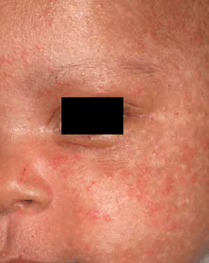
Figure 2. Miliaria associated with mild acneiform changes
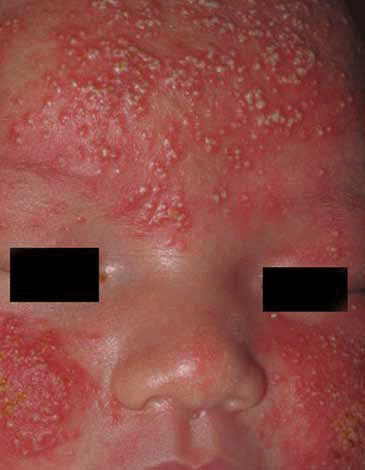
Figure 3. Pityrosporum folliculitis in a well neonate
Neonatal cephalic pustulosis is a form of pityrosporum (malessezia) folliculitis (Figure 3). Persistent papulopustules are found on the chin, cheeks and forehead. The pityrosporum yeast may relate to seborrhoeic dermatitis and pityriasis versicolor. It may stimulate acne-like follicular occlusion in children.7 Neonatal cephalic pustulosis responds promptly to topical antifungals, but these should not be used indiscreetly in the first few weeks of life given the increased absorption and toxicity risks to neonates. If used for compelling social reasons at the end of the first month of life, courses should be sparing and generally only for a few days duration. Pityrosporum pustules can also appear in a clinically nonfollicular pattern.8 Infantile acne (Figure 4), possibly related to maternal androgenic hormones, may present in the neonatal period or in later infancy with comedones and papulopustules. Severe or persistent nodulocystic cases may warrant specialist therapy and investigation for hyperandrogenism.
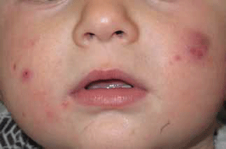
Figure 4. Infantile acne with comedones, papules, pustules and cysts
Neonatal eosinophilic folliculitis, which is distinct from the adult and HIV associated forms, may be a presentation of hyperimmunoglobulinaemia. Seen primarily on the head and neck, it can persist and recur, but idiopathic cases usually subside by 3 years of age. Older children may benefit from the application of topical steroids, and a regimen of antihistamines and occasionally dapsone, but these are not suitable early in life.
Scabies in neonates can be extensive and not evidently pruritic.
Changes may be prominent in acral sites and genitalia, similar to scabies in older children, but may also be more widespread and involve the scalp and face, with vesicles, pustules, burrows, eczema and secondary impetiginisation. Both 5% permethrin (8 hours overnight) and 5% sulphur in paraffin (twice daily for 1 week) can be used for treatment. Acropustulosis (Figure 5) is a condition thought to follow scabies, with recurrent crops of itchy vesiculopustules on the palms, soles and dorsal acral skin. It responds to topical steroids and usually settles by 2 years of age. The prescription of dapsone is rarely justified.
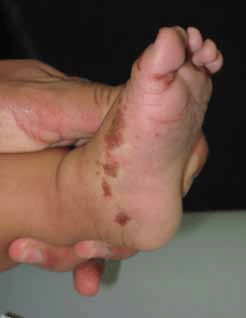
Figure 5. Acropustulosis with resolving papules and pustules
Linearly arranged vesiculopustules, particularly on the limbs and trunk, may be incontinentia pigmenti (Figure 6). This x-linked dominant condition, which usually affects females, may be associated with alopecia, eye, dental and neurodevelopmental complications. Other skin changes include linear and whorled pigmentation, warty plaques and linear atrophic areas that may follow developmental (Blaschkoid) lines.
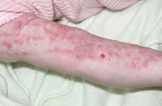
Figure 6. Linear papules and pustules of incontinentia pigmenti stage 1
Milia may be misdiagnosed as pustules, but are in fact firm 1–2 mm keratin cysts often clustered on the forehead, nose, cheeks and chin. They usually spontaneously resolve by 3–4 weeks of age. If widespread and persistent they may indicate an underlying syndrome such as trichodysplasia or epidermolysis bullosa.
Epidermolysis bullosa (Figure 7) is a mechanobullous disorder associated with various genetic mutations to skin proteins and is characterised by fragile skin, blisters and ulcers. Relatively minor trauma results in blistering and erosions. The extent of mucoutaneous, nail, dental and systemic involvement (such as oesophageal stenosis, eye, bowel, cardiac and renal disease) depends on the subtype, the junctional and dermolytic forms being more severe and sometimes lethal. Children with clinically significant mechanobullous disease should be assessed by a specialist with expertise in this area, as there are many current developments both in diagnostic testing and the treatments available. Dedicated epidermolysis bullosa nurses are now being appointed in New South Wales, Queensland and Victoria, working with paediatric dermatologists and the National Dystrophic Epidermolysis Bullosa Research Association of Australia (DebRA) to rapidly diagnose and manage these patients.
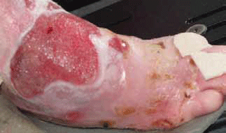
Figure 7. Blisters and ulcers of recessive dystrophic epidermolysis bullosa
Ecchymoses and crusts
Similar infective, traumatic and genetic causes should be considered in neonates presenting with ecchymoses and crusts. Coagulopathies (deficiencies of protein C or S, fibrinogen or secondary deficiencies from infection or maternal antibodies), neonatal Behcet disease, neonatal lupus and ulcerated tumours should also be considered.9 Neonatal lupus usually presents with red annular macules and plaques in neonates, often on the face. Related to maternal Ro and La antibodies, it is important to pick because of a risk of associated heart block.
Neonatal papules and Nodules
Midline nodules may represent dysembrogenesis and this must be excluded before any intervention. Pathologies include:
- head: encephalomeningocoeles, dermoid cysts, nasal glioma
- neck: thyroglossal cyst, branchial cysts
- neck and back: meningocoele and spinal dysraphism
- umbilicus: omphalomesenteric dysraphism.10
Scattered bluish nodules and papules are seen in the 'blueberry muffin baby' and may represent an underlying tumour (eg. neuroblastoma, rhabdomyosarcoma, leukaemia), infection (toxoplasmosis, rubella, cytomegalovirus, coxsackie B2, parvovirus B19, syphilis), and other extramedullary erythropoeisis (hereditary and immune triggered haemolysis).
Other cutaneous clinical clues in diagnosing nodules and plaques include Darier's sign (urtication or blistering with rubbing most often seen in mastocytosis, Figure 8), orange colour (infantile xanthogranuloma, Figure 9), red-blue colour with peripheral vessels and consistent Doppler findings (haemangioma), subcutaneous inflammation (subcutaneous fat necrosis), deep location and firm texture (fibromatoses, myfibromatoses, sclerema neonatorum), purpura and seborrhoeic dermatitis-like changes (histiocytosis). Clinical signs and imaging findings can err and tissue diagnosis may be required. Potential complications must also be identified (eg. hypercalcaemia in fat necrosis, systemic disease).
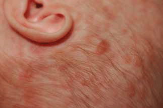
Figure 8. Urticating and blistering papules and plaques in urticaria pigmentosa (a form of mastocytosis)
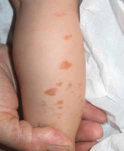
Figure 9. Infantile xanthogranuloma
Cradle cap and red scaly rash
Cradle cap derives early on from vernix, and subsequently, possibly also from sebaceous secretions. Malessezia may also contribute to cradle cap. It generally self remits, but the application of gentle baby shampoo or mineral oil (liquid paraffin) may help it soften. If there is more extensive seborrhoeic dermatitis (greasy, nonpruritic, erythematous changes of face, ears, neck, nappy area), there is a limited role for the sparing use of topical hydrocortisone and an imidazole cream.
Seborrhoeic dermatitis generally arises after 1 month, and atopic dermatitis after 2–3 months. The former is usually not itchy, whereas the latter is often characterised by considerable pruritus.
Congenital widespread desquamation with or without redness (erythroderma) raises the possibilities of ichthyosis (eg. Netherton, Sjögren-Larssen) and immunodeficiency (eg. Omenn syndrome).11 However, causes of erythroderma in the first month of life should additionally include psoriasis, pityriasis rubra pilaris, metabolic conditions (zinc deficiency, protein malnutrition, multiple carboxylase deficiency, urea cycle disorders, essential fatty acid and biotin deficiencies), scalded skin syndrome, candida and drugs.12 Zinc deficiency causes well defined psoriasiform plaques of the hands, feet, perineum, shoulders, perioral fissuring, thin hair and irritability. It usually results from inadequate zinc intake, but occasionally may result from malabsorption. Measuring zinc levels may not be reliable, but skin changes rapidly reverse with zinc replacement.
Collodion baby refers to babies encased in a tight, yellow, shiny film at birth. Although usually resulting from lamellar ichthyosis and nonbullous congenital erythroderma, it can be related to other ichthyosis syndromes such as trichothiodystrophy and possibly metabolic disease (Gaucher type 2, hypothyroidism). Thermal and fluid imbalances, infection, respiratory compromise, ectropion, feeding problems and constriction bands are all possible complications. Consequently, neonatal intensive and multidisciplinary care is required for most cases of collodion baby. Congenital erythroderma and collodion babies require specialist care.
Key points
- Minimise the use of topical toxins and unnecessary drugs in neonates.
- Consider bacterial, viral, and fungal infections in neonates presenting with pustular and vesiculobullous dermatoses.
- Simple tests such as smears and fluid culture can be very helpful.
- Nodules, bruising and crusts may all indicate serious underlying diseases.
- Midline defects may be associated with neural tube anomalies.
- Early desquamation and erythroderma may signify genetic diseases such as ichthyosis and primary immunodeficiency.
Conflict of interest: none declared.
References