The diagnosis of maxillofacial fractures can be challenging, as haematoma and swelling can mask the extent of the underlying injury. Overlooking a fracture may not have immediate consequences, but can result in disfigurement and permanent disability. Not only does this result in a disgruntled patient, it may affect their ability to continue to perform their occupation.1
Unfortunately, the incidence of maxillofacial trauma is increasing at an alarming rate. Maxillofacial trauma presentations in 2011 at the Royal Brisbane Hospital (Queensland) have risen 28% in the same 10 month period compared to 2010.2 Despite the decrease in facial trauma from motor vehicle accidents due to safety improvements such as airbags and seat belts, injuries due to interpersonal violence continue to rise. In Australia, maxillofacial fractures are the third most common trauma in falls in the elderly after neck of femur and upper limb fractures.3
Initial presentation
Weekends, night time and Monday mornings are the most common times that facial injuries present. Although some injuries may appear dramatic, they should not distract the GP from their initial assessment and management. Assessment should initially follow standard Advanced Trauma Life Support (ATLS) guidelines.4 Airway compromise, major haemorrhage and visual loss are the key problems to rule out on initial assessment. Accurate assessment is crucial.
Head trauma with involvement of the neurocranium may lead to unconsciousness, amnesia, nausea, post-traumatic headache or dizziness. The severity of this can be assessed using the Glasgow Coma Score (GCS).1 Injuries affecting the GCS need immediate referral to a hospital emergency department.
Soft tissue injuries
Disruption to the soft tissues of the face can be disfiguring and may significantly damage underlying anatomical structures. The lacrimal ducts, parotid ducts, main vascular architecture and nerves, especially the facial nerves should all be assessed for potential damage. Foreign bodies and debris should be removed with lavage, avoiding excessive debridement if possible.
The face has an excellent vascular supply. Although this can be problematic with regards to acute haemorrhage, it contributes to optimum wound healing. Optimum results are obtained with precise primary repair. However, if bleeding is difficult to control or in large wounds, the combination of immediate approximation from well positioned sutures with early referral for wound revision is appropriate.
Nasal fractures
Nasal fractures are the most common facial fracture accounting for up to 58.6% of all facial fractures.5 The main cause is interpersonal violence. Oedema may mask deformation of nasal bones. Therefore, manual assessment and palpation of the nasal base is mandatory, although this is often painful. Nonreduced nasal fractures typically lead to a flat or curved base with remaining disfigurement for the patient (‘rugby nose’).
Clinical assessment
The nasal bones should be assessed for asymmetry and mobility. Assessment of the pharynx is necessary to ensure there is no posterior nasal bleeding. Beware patients on anticoagulant therapy. A nasal speculum can help localise haemorrhage or haematoma, especially adjacent to the nasal septum.4 Septal haematoma may strip the septal cartilage of blood supply and progress to abscess formation or later cartilage necrosis, resulting in significant nasal deformity and septal perforation. An overlooked septal haematoma may critically disfigure the patient and it should always be ruled out.
Epistaxis can be alarming, but is usually controlled with the following conservative measures:
- pinching the nasal ala against the septum
- topical vasoconstrictors (nasal decongestant)
- chemical cautery with silver nitrate.6
If these measures fail, an epistaxis tampon is the next step.
Imaging
Clinical examination is usually sufficient and plain X-rays are generally of little benefit. If the nasal fracture appears to be part of a wider fracture pattern, including the frontal bone or maxilla, a computed tomography (CT) scan of the facial bones is advised.
Management
Nasal fractures usually require referral to a maxillofacial or an ear, nose and throat (ENT) unit. However, they can generally be left 5–7 days to allow the oedema to resolve, facilitating precise operative intervention. In the acute setting, an attempt can be made to grossly reduce the fracture within 4 hours of the incident.7
Mandibular fractures
Mandibular disruptions are commonly encountered. Because, unlike the rest of the facial skeleton, the mandible is a mobile bone, these injuries are very painful. Mandibular fractures are generally easy to diagnose. Patients usually report malocclusion and pain over the fracture site.
The mandible will usually fracture in two places. This is usually the site of direct impact and a fracture in an area opposite this site. This second fracture commonly involves the mandibular condyle or mandibular angle on the contralateral side (indirect fracture).1 If the impact hits the middle of the mandible (symphysis), additional indirect fractures of both condyles are common.
The mandibular condyles must be carefully assessed when a patient presents with a blow or laceration to the chin. Have a high index of suspicion of condylar fractures in children who have fallen, as these injuries are often missed and can result in lifelong pain and disability. Because of pain and discomfort mandibular fractures should be referred within 24 hours.
Clinical assessment
The initial oral inspection should include locating missing teeth, obvious fracture sites and any intra-oral laceration. The lower dental arch should be evaluated to ensure it remains intact. As with maxillary fractures, all missing teeth should be accounted for which may require radiographic evaluation of the chest. Complete disruption of the mandible and the subsequent loss of dental alignment can mimic a missing tooth.
Post-traumatic malocclusion is often reported by the patient and should generally be visible by intra-oral inspection. Test occlusion by asking the patient to close their mouth to identify any malalignment of the teeth. A step in the occlusal plane with a ruptured gingiva at the site (Figure 1) or a sublingual haematoma (Figure 2) are strong indicators of mandibular fracture.8
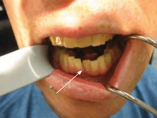
Figure 1. A step in the occlusal plane and ruptured gingival (arrow) associated with a mandibular fracture
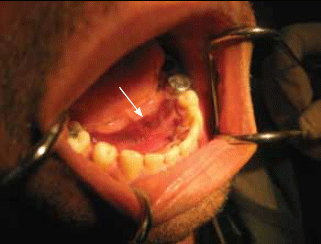
Figure 2. A sublingual haematoma (arrow) can be an indicator of a mandibular fracture
A new malocclusion, either on a single side or anteriorly, is associated with a condylar fracture. Bimanual assessment of a suspected mandibular fracture is undertaken by grasping the mandible either side of a suspected fracture site and carefully testing mobility. Pressing on both mandibular angles extra-orally and bending the midsection of the jaw may also cause pain at the fracture site.1
The mandibular nerve, as it courses through the mandibular angle or body, is often involved with the fracture. This will display itself as a parathesia or anaesthesia of the lower lip and chin on the ipsilateral side.
Imaging
Plain radiographs are first line: an orthopantomogram (OPG) and PA mandible radiograph. Computed tomography can be used if clinical signs suggest a fracture but plain X-ray appears normal.9Computed tomography is not required in the majority of cases. Additional condyle fractures should be always suspected if a fracture of the mandibular body is present.
Visualised or self reported malocclusion is the key to diagnosis. Mental nerve paraesthesia is often associated with displaced fractures. A child presenting with a laceration to the chin point and pain over the preauricular area should always be suspected as having a condylar fracture, an OPG and PA mandible radiographs are mandatory.
Management
Stabilisation of the fracture is important to minimise pain and discomfort. A soft cervical collar is recommended for stabilisation, not the traditional barrel bandage. Treatment should ideally be performed in the first 24 hours to avoid infection of the fracture site. Abscess and severe osteomyelitis are not uncommon in overlooked, untreated mandibular fractures. Prophylactic antibiotics are recommended if treatment is delayed. Penicillin, or alternatively clindamycin, are appropriate because of the disruption of the mucosa with direct communication to the underlying fracture area.10
Definitive treatment
It is beyond the scope of this article to discuss the intricacies of definitive surgical treatment. However, it is important to dispel community perception that management will involve ‘their jaws being wired shut’. Interdental wiring is now rare with few specific indications. The vast majority of these fractures are treated with open reduction and internal fixation with titanium miniplates (Figure 3). Endoscopic access surgery is increasingly popular with Australian maxillofacial surgeons, especially in mandibular condyle fractures. Condylar fractures in children are often treated conservatively with a soft diet and careful monitoring and exercises during the healing period.
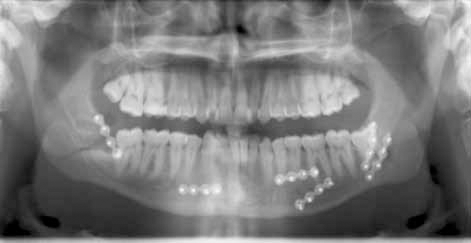
Figure 3. Multiple titanium miniplates in situ postopen reduction and internal fixation surgery
Orbitozygomatic fractures
To understand the characteristic patterns of midfacial and orbitozygomatic fractures the relevant anatomy of the facial skeleton is important as typical lines of weakness are present.
The orbit is an integral part of the zygomatic complex. The vast majority of zygomatic fractures involve the orbit, apart from the isolated zygomatic arch fracture (Figure 4).
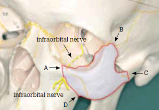
Figure 4. Zygomatic-orbital segment that is displaced in cheek bone fractures (light blue). Note that the orbital floor is always involved and usually compromises the infraorbital nerve. Discontinuities in the bone can be radiographically detected in the following areas: A) infraorbital rim; B) frontozygomatico suture; C) zygomatic arch; D) zygomaticoalveolar buttress
The initial assessment of these injuries must include an eye examination. At a minimum this examination should include visual acuity, pupillary light reflexes and ocular movements.
Any acute decrease in visual acuity should immediately be referred to an ophthalmologist or maxillofacial surgeon. The underlying cause could be a retrobulbar haemorrhage, which requires urgent treatment to avoid permanent blindness.
Clinical investigation
Common findings in the assessment of orbitozygomatic injuries include:
- palpable step at the infraorbital margin or the lateral brow area. This may coincide with the zygomatic-frontal suture
- infraorbital nerve paraesthesia. A weakness in the facial skeleton occurs where this nerve courses through the infraorbital foramen. The paraesthesia involves the ipsilateral cheek, lateral surface of nose, ipsilateral lip, upper incisor teeth and associated gingival tissue
- depression of the malar eminence (cheekbone.) A zygomatic complex fracture is normally impacted into the facial skeleton (Figure 5). This is assessed by standing behind the patient, placing one index finger on each malar eminence and comparing for symmetry. The affected side is usually more depressed
- diplopia is common due to orbital involvement. Impaired upward gaze with resultant diplopia is usually a result of oedema and haemorrhage of the periorbital tissues. Occasionally, this may be due to rectus muscle entrapment with a concomitant orbital floor fracture
- impaired mouth opening or closure may occur if the zygomatic arch is depressed and affects the underlying temporalis muscle and its insertion at the coronoid process of the mandible
- if the patient has blown the nose after the trauma they may report an immediate increase in eyelid swelling. In this case air is blown from the maxillary sinus via the fracture gaps into the orbit. Air crepitus in the periorbital soft tissue is palpable. Patients with suspected orbital fracture should not blow their nose.
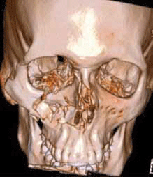
Figure 5. 3–D reconstruction of a right comminuted zygomatic complex fracture
Just as a haematoma under the tongue can be an indicator for a fractured mandible, a subconjunctival haematoma may indicate an orbitozygomatic fracture (Figure 6). If present, fracture should be ruled out radiographically.
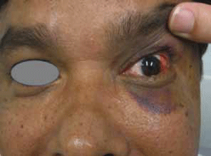
Figure 6. Left sided subconjunctival haematoma and mild periorbital haematoma that might be indicators for orbitozygomatic complex or orbital wall fracture. These haematomas always require further radiographic examination as fractures need to be ruled out
Imaging
The gold standard in investigating these injuries is a CT scan with axial and coronal views (Figure 7). We would recommend no greater than 3 mm slices. Plain facial radiographs often provide no additional information and can be falsely negative.
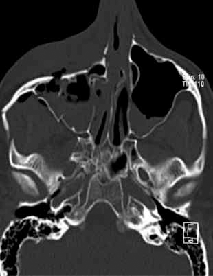
Figure 7. Axial CT of a comminuted zygomatic complex fracture
Zygomatic complex fractures often involve the orbit; therefore assessment of the eye is mandatory.
Treatment
In most instances, immediate referral is unnecessary and it is reasonable to postpone referral for 4–7 days. There are some important exceptions and these will be discussed later.
These injuries are usually treated by accurate reduction with internal fixation with miniplates and screws (Figure 8).
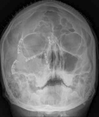
Figure 8. A zygomatic complex fracture treated with titanium miniplates and screws
Incisions are usually cosmetically hidden within the hairline, eyebrow, lower lid creases, the conjunctiva and intra-orally. Large areas of the facial skeleton can be accessed and treated with very little long term cosmetic evidence (Figure 9a, b).
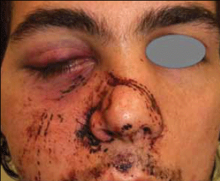
Figure 9a. A zygomatic complex and nasal bone fracture on the patient´s right side after a sporting injury
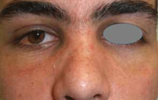
Figure 9b. The same patient after maxillofacial intervention and correct anatomical reposition and fixation of the segments. Note: the incision scars are ‘hiding’ in the folds of the eyelids leading to an excellent cosmetic and functional result
Isolated orbital fractures
The medial and inferior walls of the orbital skeleton are exquisitely thin (Figure 10). These areas are commonly involved in zygomatic complex fractures but can also occur as an isolated injury. A direct blow to the orbit or orbital rim is usually required to sustain this fracture type.
Assessment of the eye is paramount when these injuries occur.
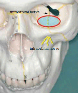
Figure 10. Anatomy of the left orbit. Area of weakness in the orbital floor highlighted. Note the passage of the infraorbital nerve through the same area
Clinical assessment
- Subconjunctival and periorbital haematoma are common
- Diplopia commonly occurs when the orbital walls are fractured (usually the floor) compromising the orbital muscles and soft tissue, sometimes entrapping them.
- Infraorbital paraesthesia or anaesthesia can occur when a fracture of the orbital floor affects the infraorbital nerve.
Imaging
Computed tomography scanning with both axial and coronal views (Figure 11) is required, although studies with newer magnetic resonance imaging (MRI) modalities suggest that in the future this may be the investigation of choice.11
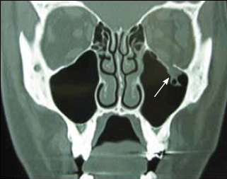
Figure 11. Coronal CT of a left orbital floor fracture (arrow)
Management
Unless there is any disturbance in visual acuity, these injuries can generally be left 7–14 days without intervention. Again this type of fracture can rarely be associated with a retrobulbar haematoma – a sight-threatening emergency.
Not all orbital fractures require repair. Some are treated conservatively. Specific parameters are followed to determine which fractures require surgical intervention.
As with orbitozygomatic fractures, a fracture of the orbit allows communication to either the maxillary, ethmoid or frontal sinuses. It is important that on presentation the patient be advised not to blow their nose or valsalva as this can produce acute facial emphysema that can be quite distressing. Similarly, air travel is not recommended for 2 weeks after this type of injury.
It is reasonable considering the antral involvement of these fractures to commence a course of antibiotic therapy: 5–7 days of amoxycillin and clavulanic acid.
Vision must be assessed, and if intact, referral can usually be delayed for 7–14 days.
Maxillary and midface fractures
Midface fractures typically run along bilateral lines of weakness in the midfacial skeleton. They may involve the maxilla, the midcentral midface or the entire midface. The latter two involve the orbits and lead to typical bilateral haematoma (‘raccoon sign’).
Clinical investigation
Midface fractures are characterised by symmetrical facial swelling, bilateral periorbital ecchymosis and bilateral subconjunctival/ periorbital haemorrhages (raccoon signs) with flattening and elongation of the midface. Mobility of the maxilla is tested by stabilising the patient’s head by applying pressure over the forehead using one hand. The thumb and forefinger of the other hand grasp the anterior maxillary ridge and pressure is then used to elicit maxillary mobility. Patients will often complain of an inability to find occlusion with the teeth and mobility of the top jaw. Bilateral infraorbital nerve paraesthesia can also occur. Later presentations may complain of continued blood clots in the posterior pharyngeal region, as the initial clotting in the maxillary antrum is cleared into the posterior pharynx.
These injuries are dramatic and painful due to maxillary mobility and require immediate referral.
Imaging
All suspected cases of midface fracture require comprehensive CT examination, particularly with orbital involvement. If further head injuries are suspected or neurological symptoms are present, the CT scan should include soft tissue and brain. Plain radiographs such as OPG, nasal sinus (occipitomental projection) and axial zygomatic arch (submentovertical projection) can be useful where CT imaging is not accessible.
In children, midfacial fractures are rarer as sinus development is incomplete. However, CT scans are indicated if there are clinical indicators such as diplopia, change in occlusion, infraorbital nerve parathesia or severe facial bruising.
In maxillary fractures occlusion and midface mobility should be checked.
Frontal bone fractures
Some individuals have well pneumatised facial sinuses predisposing them to these types of injuries. The drainage of the frontal sinus is extremely poor and assessment always involves the frontonasal ostea. The most common frontal bone fracture is an anterior table fracture (Figure 12). Most patients are initially concerned with the cosmetic deficit produced by this type of fracture (Figure 13).
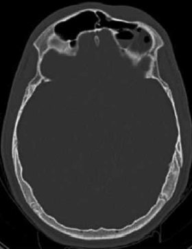
Figure 12. Depressed anterior table frontal sinus fracture
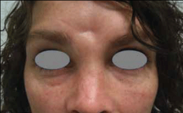
Figure 13. Obvious cosmetic deformity associated with frontal sinus fracture
Posterior table fractures are more significant but thankfully less common. These fractures involve the cranial cavity and often show entrapped air inside the neurocranium therefore requiring joint management by maxillofacial and neurosurgical teams.
Clinical assessment
Deformity can usually be visualised but must be palpated. The nasal complex can be involved and requires assessment.
Imaging
If these fractures are suspected CT is the imaging modality of choice.
Management
Anterior table fractures do not require immediate referral and can be referred up to 4–7 days postinjury. If a posterior table fracture is suspected the patient should immediately be transferred to a major hospital.
A common presentation is a cosmetic indentation to the forehead (Figure 13). This is usually only apparent acutely. Oedema and swelling will make the area appear to resolve, though the deformity will return when the swelling subsides.
Panfacial fractures
These fractures are a combination of all the previously mentioned fractures. They are unlikely to be a common presentation to general practice. As with all serious injuries, initial assessment and management should be conducted by ATLS guidelines. Due to the serious nature of these injuries and the force involved, cervical spine injury, cerebral injury and cerebrospinal fluid leak may occur. The average general practice is not equipped to deal with these types of injuries and swift transfer to a hospital should be arranged.
Vital symptoms/signs/conditions not to be missed
Although most maxillofacial injuries do not require immediate referral to a maxillofacial unit, there are some important exceptions, mostly involving the eye and vision. We stress that in assessing any maxillofacial injury that normal ATLS principles should be adhered to and this should also include a full visual assessment.
Retrobulbar haemorrhage
Retrobulbar haemorrhage is an acute condition following orbital trauma. It is due to an intraconal haemorrhage, which in turn causes a compartment syndrome of the orbit. Signs include proptosis, chemosis, ophthalmoplegia and a loss of visual acuity (Figure 14).
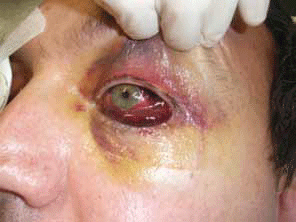
Figure 14. Early retrobulbar haematoma with loss of vision. Retrobulbar haematoma requires immediate treatment. The globe is firm to palpation
If progressive, retrobulbar haemorrhage is an emergency requiring immediate decompression. It can result in irreversible ischaemia in less than 2 hours.12–14 Refer immediately to an emergency department for management by an ophthalmologist or maxillofacial surgeon.
Proptosis, chemosis, ophthalmoplegia and loss of visual acuity is a sight-threatening emergency and requires immediate treatment.
Traumatic optic neuropathy
Traumatic optic neuropathy is an acute injury of the optic nerve with disruption of visual function.15 It is more commonly associated with maxillofacial injuries but can occur from a seemingly trivial injury. The nerve can be directly or indirectly injured. Indirect optic neuropathy is when the injury to the nerve results from the nonpenetrating effects of trauma, including haemorrhage, oedema and concussion.15
The patient has decreased visual acuity and possibly decreased pupillary reflexes. Most cases of visual loss are immediate but 10% of patients have delayed visual loss.16 Immediate referral is required as surgery in some instances can decompress the involved nerve.
White eye blowout fractures
White eye blowout fractures of the orbit (also termed ‘trapdoor fractures’) are a poorly recognised entity, resulting in delayed management and poor outcomes for patients.17 These fractures typically occur in young patients and may not be clinically obvious. The pathognomonic feature of this injury is painful restriction of eye movement (Figure 15) with nausea and occasional vomiting.17 The autonomic symptoms are likely the result of raised vagal tone secondary to the entrapment of soft tissue in the fracture line.18 Significant diplopia with restriction of upward gaze can occur. Importantly there is no subconjunctival haematoma.
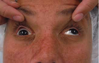
Figure 15. White eye blowout or trapdoor fracture on the patient´s right. Note the severe restriction in upward gaze of eye on right
It involves a fracture of the orbital floor with ischaemic impingement of the ocular contents in this localised area. These fractures are typically visualised on CT scan as an undisplaced linear fracture (Figure 16).
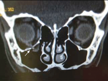
Figure 16. CT findings in a right sided white eye blowout fracture with entrapment of orbital tissue (arrow)
These injuries must be treated within 48 hours otherwise permanent restriction of ocular motility may occur.
Diplopia, nausea, vomiting with no subconjunctival haematoma in a young patient requires immediate referral.
Summary
Maxillofacial injuries are unfortunately becoming a more common presentation to general practice. The ATLS guidelines should be adhered to in the initial management of these injuries. This should be followed by a thorough and comprehensive assessment of vision and facial skeleton mobility, with careful documentation.
Practitioners should be aware of retrobulbar haemorrhage, traumatic optic neuropathy and white eye blowout fracture. Missing these injuries can have devastating consequences for the patient and medicolegal ramifications for the practitioner. If these types of injuries are suspected, immediate transfer to hospital is mandatory. Although uncommon, any patient with a panfacial fracture should also be transferred to hospital immediately.
Pain and discomfort dictate that mandibular and midface fractures should be referred within 24 hours. All other nonlife or nonsight threatening injuries can be referred within 7 days.