Nationally and internationally, less than 5% of medical undergraduate education addresses musculoskeletal conditions.3,4 Graduates may therefore lack the necessary skills to accurately assess, diagnose and manage common shoulder conditions. Around 95% of people with shoulder pain are treated in primary care settings.5 A systematic, structured approach to the assessment of shoulder conditions is essential to formulate a correct diagnosis and management plan.6,7
The human shoulder
When assessing shoulder injuries it is important for the clinician to have a sound knowledge of anatomy. Shoulder movements, stability and range of movement depend on four separate joints (Figure 1):
- sternoclavicular
- acromioclavicular
- glenohumeral
- scapulothoracic
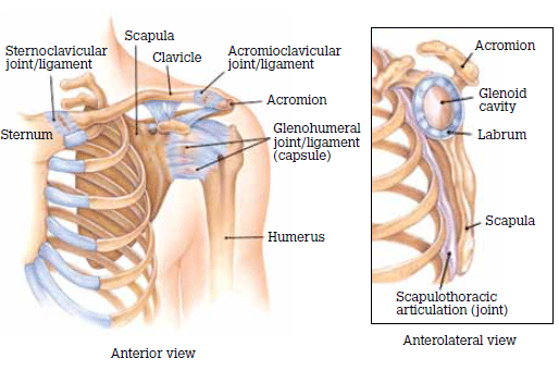
Figure 1. Basic shoulder anatomy
The glenohumeral joint is highly mobile with a trade-off of poor stability, hence integrity of the static and dynamic shoulder stabilisers is essential. The static stabilisers act independently of the shoulder position; for the glenohumeral joint they are the articulation of the joint surface and the capsulolabral complex. Dynamic stability is stability for movement in different positions and is assisted by the rotator cuff muscles and the scapular rotators including trapezius, serratus anterior, rhomboids and levator scapulae.
A general approach to assessing the injured shoulder
A focused history is the most valuable yet least effectively used tool in clinical medicine.6 A poor history and physical examination may lead to inappropriate diagnostic testing and may also influence patient outcomes.7 There is as much art as science when performing a focused shoulder history and examination.8 It requires a combination of sound medical and anatomical knowledge, clinical exposure and experience to have strong pattern recognition and clinical acumen. Focused assessment of other body systems, such as cardiovascular or neurological, may be clinically indicated.
A focused history
Patients usually present with a shoulder condition because they are experiencing pain, dysfunction or a combination of the two. The clinician must be satisfied they have adequately explored three domains:
- mechanism of injury
- pain
- dysfunction.
Mechanism of injury
Asking about the mechanism of any specific injury is critical, particularly about three factors relating to the time of injury: anatomical site, limb position and subjective experiences.
Take care to clarify the patient’s description of the anatomical site.9 A description of the arm position at the time of the injury is also valuable. For example, falling on an abducted and externally rotated arm increases the risk of shoulder dislocation or subluxation. Finally, exploring the subjective experiences of the patient at the time of injury can be useful. For example, a snapping or cracking sound may be related to a bone or ligament breaking; feeling something ‘pop out’ may suggest a joint dislocation or subluxation.
Pain
Pain is a subjective perception which is moulded by genetic and cultural makeup and experienced life events.10 Musculoskeletal pain is often poorly localised and may be referred. Pain may be classified in many ways and the terminology used are varied11 and continually revised. Figure 2 outlines a useful classification of pain into nociceptive and non-nociceptive. Table 1 provides a mnemonic for obtaining a pain history.
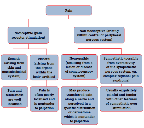
Figure 2. Pain classification
| Table 1. OLD CARTS pain mnemonic |
Onset: when did it start and how?
Location: Have the patient map it on their unaffected shoulder or on yours
Duration: How long does the pain last?
Character: A sharp, shooting pain may suggest a neuropathic cause, whereas a dull deep ache may be emanating from a joint or muscle
Aggravating and relieving factors: Pain relieved by gentle movement suggests an inflammatory cause. Pain in all directions of movement suggests joint pathology, whereas pain in one or two planes of movement may relate to muscle or tendon pathology
Radiation: Shoulder pain rarely radiates below the elbow or across the midline. Pain radiating below the elbow may be due to nerve irritation, either the cervical spine or an unstable shoulder putting traction on adjacent nerves
Timing: How long has the patient had pain and what is the natural history of this pain?
Severity: A visual analogue scale gives a baseline so pain progression or resolution may be mapped. The impact on the patient’s activities of daily living should be assessed |
Dysfunction
Clarify the degree of dysfunction, what causes the dysfunction and how this impacts on the patient (Table 2). A focused history should also screen for red flag conditions and other conditions relevant to the shoulder (Table 3).
| Table 2. Assessment of dysfunction |
- Which movements are limited? This can help isolate the structure
- Consider the following if movements are limited by:
- pain: tendinopathy, impingement, sprain/strain, labral pathology
- mechanical block: labral pathology, frozen shoulder
- night pain (lying on affected shoulder): rotator cuff pathology, anterior shoulder instability, ACJ injury, neoplasm (particularly unremitting)
- sensation of ‘clicking or clunking’: labral pathology, unstable shoulder (either anterior or multidirectional instability)
- sensation of stiffness or instability: frozen shoulder, anterior or multidirectional instability
|
| Table 3. Shoulder red flag conditions |
- Polymyalgia rheumatica. Often presents as bilateral shoulder pain and weakness. These patients must be assessed for temporal arteritis
- Acute compartment syndrome. May result from significant limb swelling following an injury or an excessively tight bandage or cast. The pain is disproportionate to the injury. Pulselessness of the limb does not usually occur, or is a very late sign. This condition is a surgical emergency
- Open fractures
- Fractures with nerve or vascular compromise
- Skin, but more particularly joint infections
- Neoplasia
- Serious and life threatening conditions that present with symptoms mimicing shoulder pain, such as referred ischaemic cardiac pain
|
A focused shoulder examination
Examination progresses through inspection, palpation and range of movement of each of the joints of the shoulder. Figure 1 demonstrates these joints. Adequate exposure of the shoulder, including the entire scapular region and at least to nipple line is essential. Table 4 outlines some key aspects of each stage of the focused examination.
| Table 4. The focused shoulder examination |
Inspection – compare both sides
Size, shape, position, scars, lumps and bumps, colour, bruises, erythema, swelling
Palpation
- Tenderness and altered sensation (subjective)
- Surface temperature, texture (objective)
- a hot tense surface may indicate infection, inflammation/synovitis, recent trauma or tumour
- Swelling?
- may indicate effusion, tumour, nodule or bone changes
- Crepitus with movement?
- occurs in osteoarthritis, tendinopathy and fracture
Movement
- Active movement
- range of movement, fluidity of movement, pain with movement
- Passive movement – difficult if the patient guards. Only necessary if active movement is limited. Can help differentiate between:
- weakness, secondary to nerve of muscle
- block from mechanical cause or due to pain
- Measurement – as baseline, to gauge progress
Special tests – as indicated (see ‘Shoulder injuries’ in this issue of AFP) Other systems examination as clinically indicated
- Cervical spine, neurological assessment of upper limb, cardiovascular assessment
|
Inspection
The key principle with this phase of the shoulder examination is symmetry. The shape, position and function of each shoulder should be relatively similar. Some differences can occur due to shoulder dominance; the dominant shoulder may sit lower and may appear somewhat larger due to larger muscle mass.
Palpation
Several key structures should be closely examined, as these are often the source of shoulder pain and dysfunction. Palpation combines objective information such as temperature and tissue firmness and subjective information from the patient such as tenderness and dysthesia. Pain is poorly localised but tenderness is well localised. Tenderness strongly suggests that the pathology may be at the site where the tenderness was elicited, although this is difficult to elicit for deep structures. Conversely, if the patient experiences pain but the site is nontender the pain may be referred.
Identify and palpate the following (Figure 3):
- sternoclavicular joint
- acromioclavicular joint. Pain from this region is often secondary to subluxation or osteoarthritis and may be poorly perceived by the patient until palpated
- long-head of bicep tendon in the bicipital groove. Proximal bicep tendinopathy is a common condition which is often missed
- greater tuberosity of the humerus – the insertion point of some rotator cuff muscles
- periscapular muscles including rhomboids
- cervical spine to assist excluding referred pain.
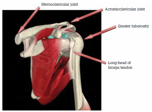
Figure 3. Anterior shoulder structures for palpation Reproduced with permission Primal Pictures. Available at www.primalpictures.com
Range of movement
The patient performs active movements in all functional planes for the shoulder. This includes flexion, extension, abduction, adduction and internal and external rotation.
Estimate the range of movement and compare the affected with the unaffected shoulder and with the normal expected range. Measuring devices such as tape measures and goniometers have poor inter- and intra-rater reliability.12–15
Special tests commonly used in shoulder assessment are covered in detail in the article ‘Shoulder injuries: management in general practice’ in this issue of Australian Family Physician.
Key points
- Musculoskeletal presentations are common in the primary care setting.
- Shoulder complaints contribute to many of these presentations, therefore a sound and confident approach to the shoulder presentation is important.
- Having a systematic and structured approach to the shoulder history and examination ensures that key aspects of the condition are elicited and important conditions are not missed.
- Information gathered in this process can help guide decisions about the need for special tests or investigations and ongoing management.
Conflict of interest: none declared.
References