Australia has good epidemiological data concerning eye disease and visual loss from two large cross-sectional, population-based studies: the Melbourne Visual Impairment Project and the Blue Mountains Eye Study, which together have provided data on nearly 9000 participants. These studies highlight an exponential increase in visual loss with increasing age: the estimated prevalence of low vision (worse than 6/12, or driving vision) and blindness (worse than 6/60) increased from 11% and 0.7%, respectively, in people aged 70–79 years, up to 39% and 17%, respectively, in those aged >90 years.11
There are numerous potential causes of visual loss; however, this article aims to highlight the areas of greatest relevance to and encountered most frequently in the elderly, namely refractive error, cataracts, age-related macular degeneration, diabetic retinopathy and glaucoma.
Refractive error
Uncorrected refractive error may be perceived as primarily a problem in the developing world, but it still remains highly relevant in developed countries. A study combining the datasets from the Melbourne Visual Impairment Project and the Blue Mountains Eye Study concluded that 62% of low vision and 4% of blindness in Australia were attributable to refractive error alone.11
Barriers to refractive correction in the elderly may be social, financial, cognitive or logistical if living in isolated rural communities or nursing homes. Refractive error can be identified in patients presenting with reduced vision by testing for improvement of visual acuity with a pinhole. Although non-correctable visual impairment has the greatest impact on individuals, visual impairment resulting from refractive error has still been shown to affect physical and social functioning.12 In a randomised controlled trial assessing the impact of immediate refractive correction on nursing home residents, Owsley et al found significant improvements in reading, psychological distress, activities and hobbies, and social interaction.13
Cataract
When vision cannot be improved by refraction, the next consideration for the elderly should be the possibility that cataracts have developed. Cataracts represent the leading cause of blindness worldwide and are extremely common with increasing age. In a cohort of the Melbourne Visual Impairment Project investigating visual impairment in elderly institutionalised Australians, the prevalence of cataract or previous need for cataract surgery increased from 56% in those aged 70–79 years to 97.5% in people aged ≥90 years.14 Age is not a barrier to consideration of cataract surgery, which should be considered for any patient who has a problem with visual function or performing activities of daily living, and wishes to improve their vision with surgical intervention. As approximately 95% of cataract surgery is performed under local anaesthetic,15 medical comorbidities are less critical than for most operations.
Medical problems of most relevance are head tremor, the inability to lie relatively flat without excessive shortness of breath, and cognitive status such that the patient is unable to remain still for about 20 minutes for the operation. If these situations arise, surgery under general anaesthetic may be offered, subject to anaesthetic fitness for surgery. Uncontrolled hypertension may also prohibit surgery, requiring liaison with the GP for optimal control of blood pressure.
A large proportion of elderly patients are on systemic anticoagulant or antiplatelet medications: a multicentre audit of 55,567 cataract operations in 2009 found that one-third of patients were taking aspirin, warfarin, clopidogrel or dipyridamole.16 No increase in potentially sight-threatening complications was identified in these patients; these medications are not prohibitive to pursuing cataract surgery.
A study assessing the impact of cataract surgery on health-related quality of life in nursing home residents found that as well as improvement in visual acuity, patients showed significant improvement in psychological distress and social interaction after undergoing cataract surgery.17
Age-related macular degeneration
Although data from the Blue Mountains Eye Study found that cataracts were the leading cause of visual impairment in the older population, age-related macular degeneration (AMD) represented the principal cause of blindness. AMD is broadly divided into two types: dry AMD and wet AMD, although the former may progress to the latter.
The clinical presentation of dry AMD is usually a gradual deterioration of vision in one or both eyes and increasing difficulty with tasks such as reading and driving. A more acute loss of central vision with metamorphopsia (distortion of straight lines) is suggestive of wet AMD. The diagnosis may be confirmed with dilated slit-lamp examination, optical coherence tomography (OCT) scans (Figure 1) and fluorescein angiography of the fundus, People with symptoms suggestive of wet AMD should be referred urgently for ophthalmic assessment as there is the potential for rapid progression and irreversible damage in wet AMD.
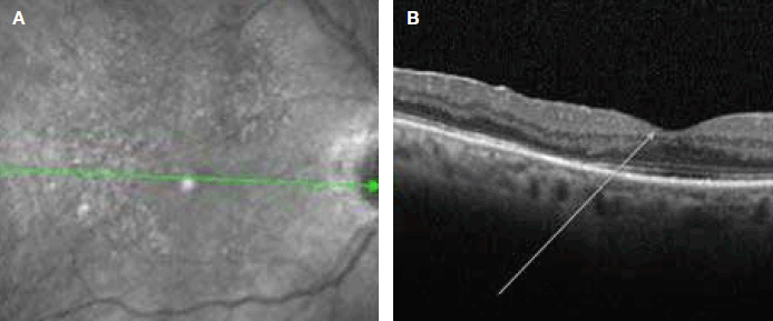
Figure 1. OCT scans
A. Arrow indicates a normal foveal contour
B. Arrow indicates the presence of sub-retinal fluid in wet AMD
Currently, there is no treatment to improve vision in dry AMD, although smokers should be advised to stop smoking to reduce the risk of progression. The AREDS study found that taking antioxidant supplements (vitamin A, vitamin C and beta carotene) combined with zinc and copper modestly reduces the risk of progression of AMD in certain subgroups of patients.18 It is known, however, that beta-carotene may increase the risk of lung cancer in smokers and the more recent AREDS2 trial, therefore, found that it may be more appropriate to substitute lutein and zeaxanthin for beta-carotene in the supplements.19
For most people with wet AMD, the treatment involves intravitreal injection of anti-vascular endothelial growth factor (VEGF) agents. These agents have revolutionised the management and visual prognosis of wet AMD over the last decade and frequently, patients’ vision will be stabilised or improved with treatment. Anti-VEGF agents include ranubizimab, which may need to be given at up to monthly intervals, or more recently, the alternative aflibercept, which can reduce the frequency of injections to every two months. The frequency of follow-up and intravitreal injections may represent significant logistical challenges for some elderly patients. Consideration is given to stopping treatment in the presence of significant visual deterioration despite optimum treatment, macular fibrosis/scarring or failure of the visual acuity to improve despite resolution of sub/intra-retinal fluid with treatment.
Despite the profound loss of central visual acuity that may result from AMD, the condition spares the peripheral retina and fortunately, therefore, many patients are able to maintain an independent lifestyle using their peripheral vision. Vision Australia and other low vision services enable patients to access advice and visual aids to assist them in their activities of daily living and maintain independence.
Diabetic retinopathy
All people with diabetes mellitus are at risk of developing diabetic retinopathy (Figure 2). Given the increasing prevalence of diabetes and an ageing population, diabetic retinopathy will affect an increasing number of elderly people. The AusDiab Study Group concluded that 15% of people with diabetes have diabetic retinopathy.20 The Australian National Health and Medical Research Council (NHMRC) recommends that all people with diabetes, including those who do not have diabetic retinopathy, should have a dilated fundal examination at least every 2 years.21 If diabetic retinopathy is detected, examinations should be performed at least annually thereafter, with greater frequency according to the severity.
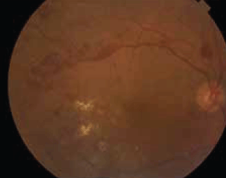
Figure 2. Diabetic retinopathy showing widespread retinal haemorrhages and hard exudates
It should be emphasised that, aside from ophthalmological treatment, good glycaemic, lipid and hypertensive control will all reduce development and progression of diabetic retinopathy22–25 and patients should liaise with their GP for optimal management of these risk factors.
In the presence of diabetic retinopathy (Figure 2) , visual loss may result from macular oedema or proliferative diabetic retinopathy, causing vitreous haemorrhage or tractional retinal detachment. Diabetic macular oedema is managed with focal laser treatment to areas of leakage and, increasingly, adjunctive intravitreal anti-VEGF agents, such as those used in wet AMD. Proliferative diabetic retinopathy is managed with pan-retinal photocoagulation laser, which reduces the angiogenic drive from ischaemic retina with subsequent regression of the new vessel growth. Vitrectomy surgery may be performed for removal of vitreous haemorrhage, relief of retinal traction and internal completion of pan-retinal laser.
Chronic open-angle glaucoma
Chronic open-angle glaucoma is an intraocular pressure (IOP)-related progressive optic neuropathy with accompanying characteristic changes in the optic nerve and visual field (Figure 3). The Blue Mountains Eye Study found a 3% prevalence of open-angle glaucoma in people aged >49 years, but an exponential rise in prevalence was observed with increasing age.26 Glaucomatous damage of the optic nerve can cause irreversible visual field loss, which, if untreated, may progress to complete blindness; the aim of treatment, therefore, is to prevent visual disability during a person’s lifetime, or progression of disability if this has occurred. Currently, the only established treatment for glaucoma is lowering the IOP and this is most commonly achieved medically; alternatives are laser (trabeculoplasty of the trabecular meshwork or cyclodiode of the ciliary body) or surgical treatment.
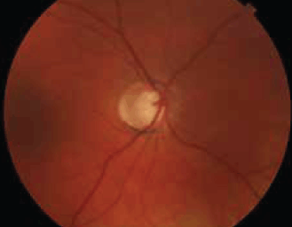
Figure 3. Advanced cupping of the optic nerve in glaucoma
When topical treatment is prescribed for the elderly, consideration should be given to the most appropriate agent. Prostaglandin analogues are the most commonly used first-line treatment. They are generally well tolerated and have few systemic side effects. Topical beta-blockers should be avoided in people with severe hypotension, obstructive airways disease, uncontrolled cardiac failure, bradycardia or second/third-degree atrioventricular block. Alpha-adrenoceptor agonists (eg. brimonidine) are unsuitable for patients with severe cardiovascular disease and may interact with monoamine oxidase inhibitors and tricyclic antidepressants. Carbonic anhydrase inhibitors (eg. brinzolamide, dorzolamide) should be used with caution in people with renal impairment. Laser treatment may be more appropriate in patients who cannot be adequately and safely treated with eyedrops, are unable to instil eyedrops, or are not compliant with treatment because of cognitive impairment.
Good communication between primary and secondary care providers is key in ensuring that treatment and management decisions are appropriate and include consideration of comorbidities and lifetime expectancy. Patients with established glaucoma almost invariably require lifelong follow-up in view of the potential for irreversible visual loss, although the frequency of review should be informed by the patient’s age, life expectancy and the extent and, critically, the rate of progression of visual field loss.
Conclusion
Many ophthalmic problems may be encountered in the elderly, but refractive error, cataract, AMD, diabetic retinopathy and chronic open-angle glaucoma represent the most common conditions and the principal causes of visual impairment in the elderly. As demographic ageing of the population occurs, the burden of these diseases will continue to rise as the number of people suffering from impaired vision and blindness increases.
Even in the absence of established pathology, elderly people should be advised to have an eye examination at least every 2 years so that eye disease, which is often asymptomatic, may be detected and treated, thereby preventing further visual loss. Many elderly people regard deteriorating vision as an inevitable sign of ageing – this is not the case and timely intervention may help prevent loss of sight and the associated negative impact on quality of life that this brings.
Acknowledgments
Jonathan Goodfellow acknowledges financial assistance from HCA International Foundation in supporting his fellowship training.
Conflict of interests: None.
Provenance and peer review: Commissioned; externally peer reviewed.