How does it work?
Bone scans use technetium-99m labelled bisphosphonates ('diphosphonates') such as Tc-99m methylene diphosphonate (MDP) and Tc-99m hydroxy diphosphonate (HDP). Like their therapeutic counterparts, these radiolabelled bisphosphonates bind to hydroxyapatite at sites of active bone formation (osteogenesis). Osteogenesis is a nonspecific response of bone to a range of stimuli, such as physiological growth/turnover, mechanical stress or injury (including fractures), infection and involvement by tumour. Other possible mechanisms of increased activity on bone scans include increased blood flow, increased mineralisation of osteoid and interrupted sympathetic nerve supply.1
In a normal bone scan, performed 2–4 hours after radiotracer injection (Figure 1), some areas appear more intense compared with the rest of the skeleton. This can be due to the configuration of the bones and their proximity to the camera, and include the iliac crests and the sacroiliac and sternoclavicular joints. The sternum often has residual ossification centres which can display increased uptake. Tracer which is not taken up by bone is excreted unchanged in the urine; hence, the kidneys and bladder are routinely seen on scans.
In addition, images can be obtained during radiotracer injection to assess regional blood flow, and within the first minute or two of the injection to assess tissue vascularity ('blood pool' activity) and capillary leakage. Flow and blood pool images are typically obtained when there is suspicion or evidence of acute inflammation due to infection (eg. active osteomyelitis) or trauma (eg. recent fracture).
As well as planar imaging, tomographic imaging (single photon emission computed tomography, SPECT), analogous to X-ray computed tomography (CT), can be undertaken. With newer devices, a low dose CT can also be acquired to produce a fused SPECT/CT image which permits precise co-registration of abnormal bone scan activity with skeletal anatomy. With or without CT fusion, the images can be reconstructed in transaxial, coronal and sagittal planes.
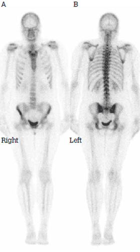
Figure 1. Normal bone scan in standard projections: A) anterior and B) posterior
When might it be helpful?
The list of potential indications for bone scans is long,2 but many of these conditions are more commonly encountered in the hospital rather than community setting. The main categories are summarised in Table 1. Figure 2 shows the typical appearance of some of these.
Table 1. Common indications for bone scan1
| Detection and follow up of metastatic disease |
| Differentiation between osteomyelitis and cellulitis |
| Determination of bone viability: infection or avascular necrosis |
| Evaluation of fractures difficult to assess on radiograph (stress fractures, fractures of complex structures and possible fractures in nonaccidental injuries of children) |
| Evaluation of prosthetic joints for infection or loosening |
| Determination of biopsy site |
| Evaluation of bone pain in patients with normal or equivocal radiographs |
| Evaluation of the significance of an incidental skeletal finding on radiographs |
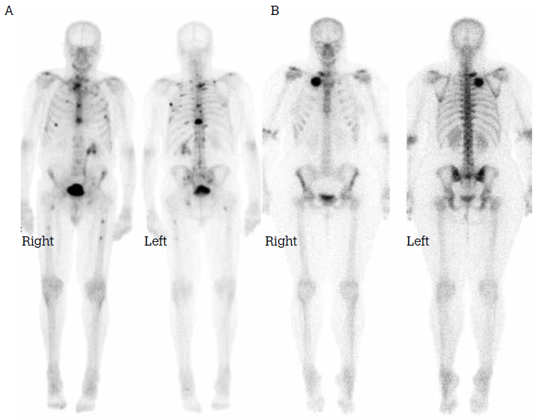
Figure 2. A) Multiple skeletal metastases; B) Fibrous dysplasia of the right first rib
When might a bone scan be positive when the X-ray is negative?
Whereas the typical radiographic features of a fracture (such as mineralisation loss and callus formation) may take several days to weeks to evolve, bone scans become abnormal within a day or two after the event as sufficient osteogenic response to the injury will have developed for detection by this method. Achieving an earlier diagnosis may in turn facilitate earlier management.
For example, a bone scan can differentiate between 'shin splints' (periostitis) and stress fractures in those who run (Figure 3). These conditions present similarly with exertional leg pain but require very different management. Shin splints typically demonstrate normal blood pool activity and linear activity on delayed scans along the length of the posteromedial tibial cortex at the insertion of the soleus muscle. Stress fractures are associated with focally increased blood pool activity and focal or fusiform uptake at the fracture site on delayed images. Conversely, a normal bone scan almost completely excludes either of these conditions.
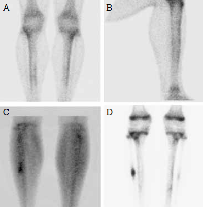
Figure 3. 'Shin splints' in (A) anteroposterior and (B) lateral views. Acute right fibular stress fracture in (C) blood pool phase and (D) delayed phase (with linear uptake at the physes indicating normal active growth plates in this adolescent); the faint abnormality in the left fibula in (D) is due to an older, healing stress fracture
How can a bone scan be helpful in nonathletic injuries?
The use of bone scans is not limited to athletic patients. Bone scans can also play an important role in the diagnosis of radiographically occult fractures (such as undisplaced femoral neck fractures) as well as sacral and vertebral insufficiency fractures (Figure 4).
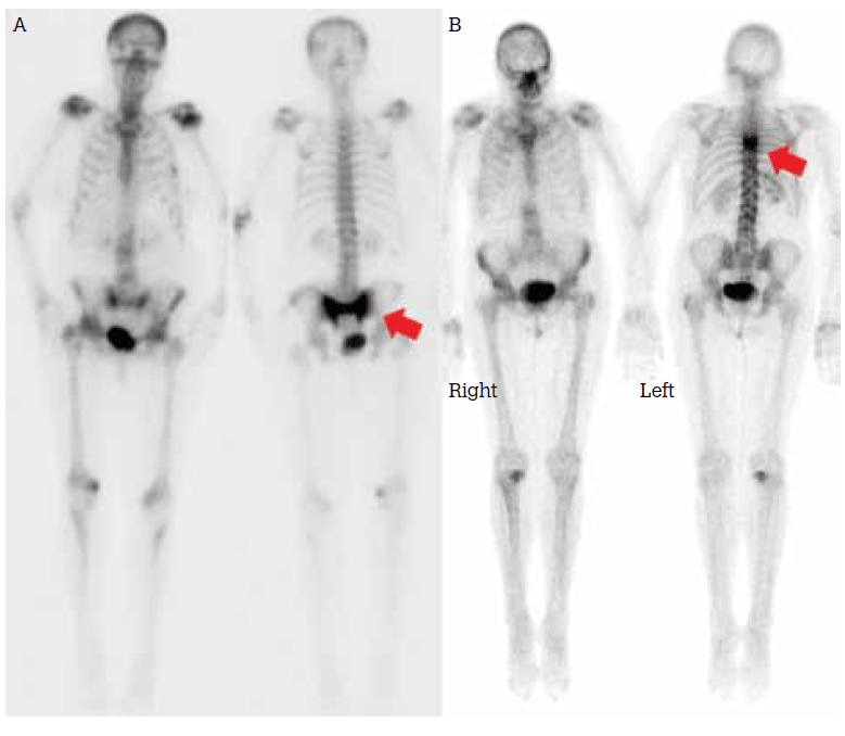
Figure 4. Some nonathletic injuries diagnosed with bone scan: A) sacral insufficiency fracture (with bilateral traumatic sacroilitis); B) vertebral insufficiency fracture
Can anything further be done if planar bone scan is inconclusive?
Tomographic (SPECT) imaging is helpful in certain situations as it provides improved spatial localisation in complex structures (such as vertebral elements). An example of this is its role in detecting pars interarticularis fractures in young patients, which may not be evident on conventional planar imaging. As noted, SPECT/CT permits functional/anatomical image co-registration.
How can it help with complications of prostheses?
Complications of hip and knee prostheses such as periprosthetic fracture, infection and loosening – all of which present with a painful prosthesis – may be distinguished with bone scan by their characteristic appearances. The first two conditions but not the last will show increased blood pool activity; the pattern of radiotracer distribution on delayed-phase images can be distinctive. Fractures show focal uptake, infections are associated with a diffuse pattern of uptake and prosthesis loosening is classically associated with uptake at the fulcra of the loose prosthesis, such as at the lesser trochanter and the tip of the femoral stem in a loosened hip prosthesis. Of particular note is the very high sensitivity of bone scan for these conditions – a negative bone scan effectively excludes these prosthetic complications.
If the bone scan suggests infection, then a labelled white cell scan should be performed for confirmation. Infection may be excluded if the bone scan is suggestive but the white cell scan is negative (Figure 5).
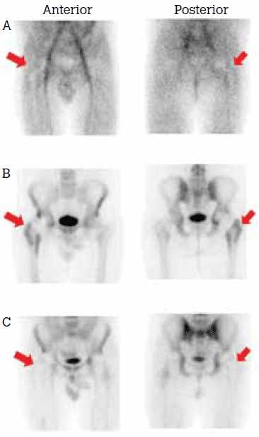
Figure 5. Increased activity associated with the hip prosthesis in (A) blood pool and (B) delayed phases. The X-rays were normal; C) Absent white cell activity excluded infection
What bone conditions is it not suitable for?
The bone scan has a low sensitivity for tumours that are confined to the marrow (such as myeloma) and those that are predominantly osteolytic with little or no osteogenic reaction (such as myeloma and renal cell carcinoma). Bone scanning is not helpful for the diagnosis of osteoporosis; bone mineral densitometry is the appropriate test for this condition.
What is the radiation dose?
Radiation dose is measured in units of millisieverts (mSv). For typical activities of Tc-99m bisphosphonates administered to adults, the effective radiation dose (an estimate of the dose to the whole body) is approximately 6.0 mSv.2 In Table 2, this is compared to the background radiation level in Australia and to the effective doses from some other commonly performed diagnostic imaging tests.
In order to reduce radiation doses to the bladder, ovaries and testes from tracer excreted in the urine, good hydration and frequent voiding are advised.
Table 2. Radiation dose for bone scan and other common diagnostic imaging tests
| Test/situation | Total effective dose (mSv) |
|---|
| Chest X-ray4 |
0.1 |
| Annual Australian background5 |
1.5–2.0 |
| Bone scan2 |
6.0 |
Sestamibi myocardial perfusion scan
(2 day rest stress protocol)1 |
11.3 |
| CT pulmonary angiogram6 |
13.7 |
| CT abdomen + pelvis4 |
15.0 |
Are there any contraindications?
Apart from a definite previous reaction to the bisphosphonate (a very rare event), there are no absolute contraindications.
Claustrophobia is a relative contra-indication. In such cases, limited views of a peripheral site may be adequate at the discretion of the nuclear medicine physician. Pregnancy is another relative contraindication.
Tc-99m labelled bisphosphonates are minimally excreted in breast milk so breastfeeding should be delayed for 1 hour after injection.3 Some patients may prefer to express breastmilk and bottlefeed for that period.
Case study 1
A man, 28 years of age, had persistent pain in the right anatomical snuffbox following a fall onto his outstretched right hand. X-ray was normal, prompting referral for a bone scan. It demonstrated focally increased radiotracer accumulation in the scaphoid on both blood pool and delayed images, typical of a scaphoid fracture (Figure 6).
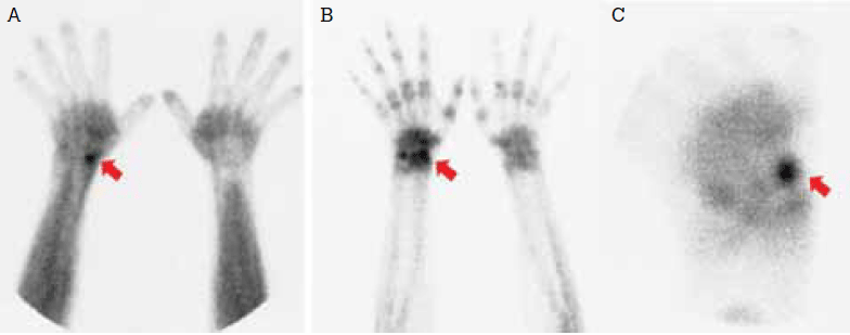
Figure 6. Scaphoid fracture with corresponding intense localised blood pool (A) and delayed phase activity (B). Magnified (pinhole) delayed image (C)
Case study 2
A man, 72 years of age, sought investigation of 3 months of chronic tiredness. He had no other specific symptoms. Blood count and biochemical profile were normal, apart from an isolated elevation in serum alkaline phosphatase level. Bone scan showed abnormal, intense radiotracer uptake in the calvarium, jaw, multiple vertebral bodies, pelvis and an expanded and bowed right femur (Figure 7). These findings are typical of polyostotic Paget disease.
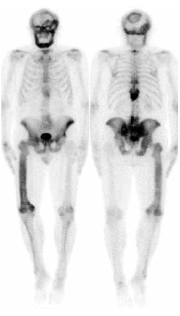
Figure 7. Polyostotic Paget disease
Conflict of interest: none declared.